
Method and device for detecting ischemic stroke focus based on brain CT image

A technology of ischemic stroke and CT images, which is applied in the field of ischemic stroke lesion detection and can solve the problem of indistinct features

Active Publication Date: 2019-03-22
BEIJING UNIV OF POSTS & TELECOMM
View PDF2 Cites 8 Cited by
  • Summary
  • Abstract
  • Description
  • Claims
  • Application Information

AI Technical Summary

Problems solved by technology

However, in the prior art, CT has not yet been used as an examination method for ischemic stroke, because the lesions of ischemic stroke are not obvious in brain CT images

Method used


Image

  • Method and device for detecting ischemic stroke focus based on brain CT image
  • Method and device for detecting ischemic stroke focus based on brain CT image
  • Method and device for detecting ischemic stroke focus based on brain CT image

Examples


Embodiment Construction

[0097] The following will clearly and completely describe the technical solutions in the embodiments of the present invention with reference to the accompanying drawings in the embodiments of the present invention. Obviously, the described embodiments are only some, not all, embodiments of the present invention. Based on the embodiments of the present invention, all other embodiments obtained by persons of ordinary skill in the art without making creative efforts belong to the protection scope of the present invention.

[0098] In order to effectively detect ischemic stroke lesions through brain CT images, embodiments of the present invention provide a method and device for detecting ischemic stroke lesions based on brain CT images.

[0099] First, a method for detecting ischemic stroke lesions based on brain CT images provided by an embodiment of the present invention will be described in detail.

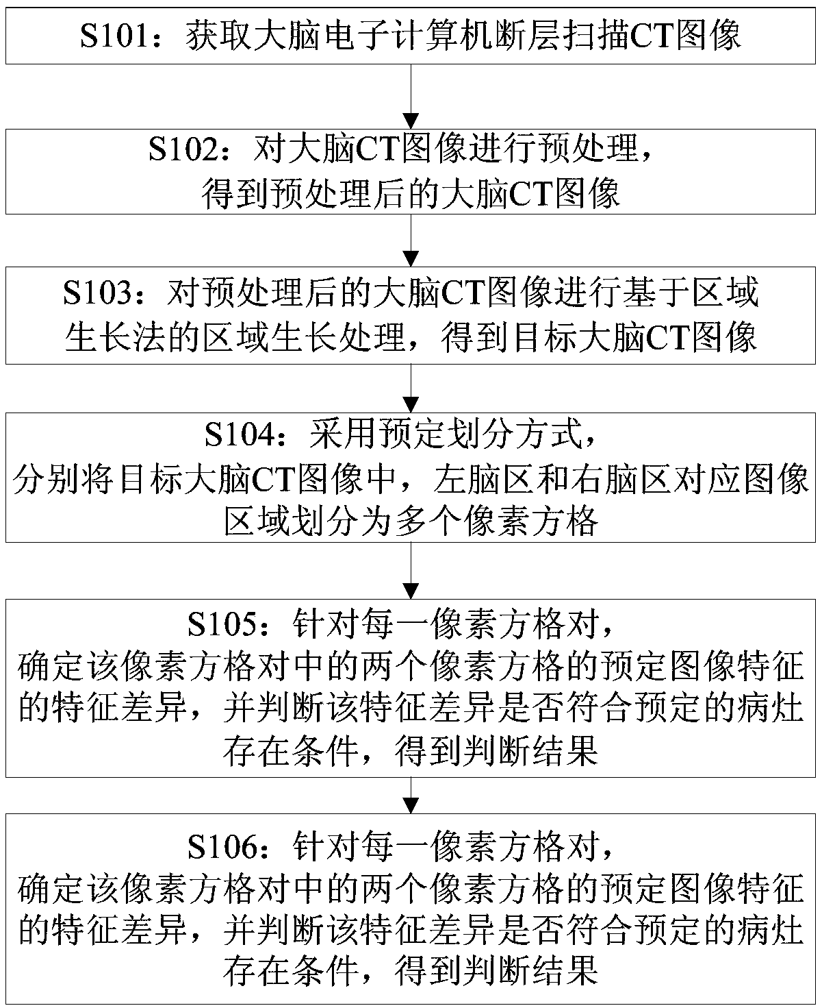
[0100] Such as figure 1 As shown, a method for detecting ischemic stroke lesi...


PUM


Abstract

The invention implements a method and a device for detecting ischemic stroke focus based on brain CT images. In this method, brain CT images were preprocessed and region growing based on region growing method, which increased the difference of image characteristics between lesion region and normal brain. Then, based on the symmetry of the brain, The images of the left and right brain regions are divided into a plurality of pixel squares, and the characteristic differences of two pixel squares with symmetry positions in the left and right brain are compared one by one, so that it is possible todetermine which pixel square contains the focus of ischemic stroke according to the characteristic differences. Through the above steps, the purpose of effectively detecting ischemic stroke lesions by brain CT images is realized.

Description

technical field [0001] The invention relates to the technical field of brain CT image detection, in particular to a method and device for detecting ischemic stroke lesions based on brain CT images. Background technique [0002] An ischemic stroke is damage to brain tissue caused by a blockage in a blood vessel that prevents blood from flowing to the brain. MRI (Magnetic Resonance Imaging, Magnetic Resonance Imaging) is often used clinically to detect ischemic stroke lesions to diagnose stroke diseases. However, the golden time window for stroke treatment is 4.5 hours after the onset of stroke, MRI scans are slow, and the cost of equipment is high, so it is not easy for general hospitals to equip them. Therefore, these shortcomings of MRI can easily lead to delays in the timing of treatment. CT (Computed Tomography, computerized tomography), as a common medical examination method in clinical practice, has the advantages of fast film production and low equipment cost. Moreo...

Claims


Application Information

Patent Timeline
IPC IPC(8): G06T7/00G06T7/11G06T7/187G06T7/73G06T5/00G06T5/20G06T5/40
CPCG06T5/20G06T5/40G06T7/0012G06T7/11G06T7/187G06T7/73G06T2207/20024G06T2207/10081G06T2207/30016G06T5/92G06T5/80
Inventor 刘军张龙祝闯杨洁刘恋杨柳缪中荣
Owner BEIJING UNIV OF POSTS & TELECOMM
Learn More