A pulmonary nodule segmentation method based on a CT image

A technology of CT images and pulmonary nodules, applied in the field of medical image processing, can solve the problems of poor robustness and low segmentation accuracy, achieve accurate separation, improve segmentation accuracy, and reduce the workload of manual judgment

Pending Publication Date: 2019-04-02
上海藤核智能科技有限公司
View PDF4 Cites 2 Cited by
  • Summary
  • Abstract
  • Description
  • Claims
  • Application Information

AI Technical Summary

Problems solved by technology

[0005] The purpose of the present invention is to provide a lung nodule segmentation method based on CT images to solve the problems of low segmentation accuracy and poor robustness in the prior art, and can accurately separate nodules and blood vessels at the same time

Method used


Image

  • A pulmonary nodule segmentation method based on a CT image
  • A pulmonary nodule segmentation method based on a CT image

Examples


Embodiment Construction

[0033] The method for segmenting pulmonary nodules based on CT images of the present invention will be described in more detail below in conjunction with schematic diagrams, wherein a preferred embodiment of the present invention is shown, and it should be understood that those skilled in the art can modify the present invention described here and still achieve Advantageous effects of the present invention. Therefore, the following description should be understood as the broad knowledge of those skilled in the art, but not as a limitation of the present invention.

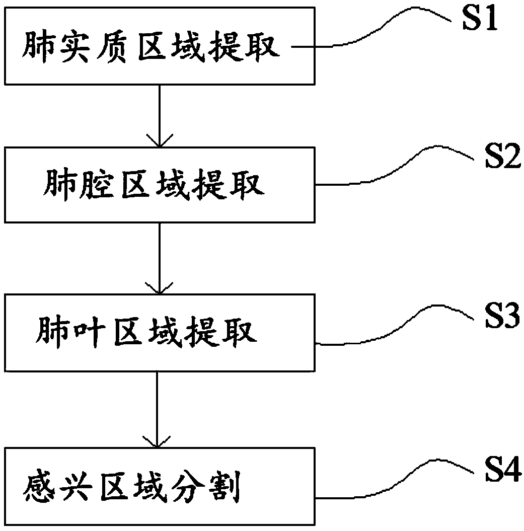
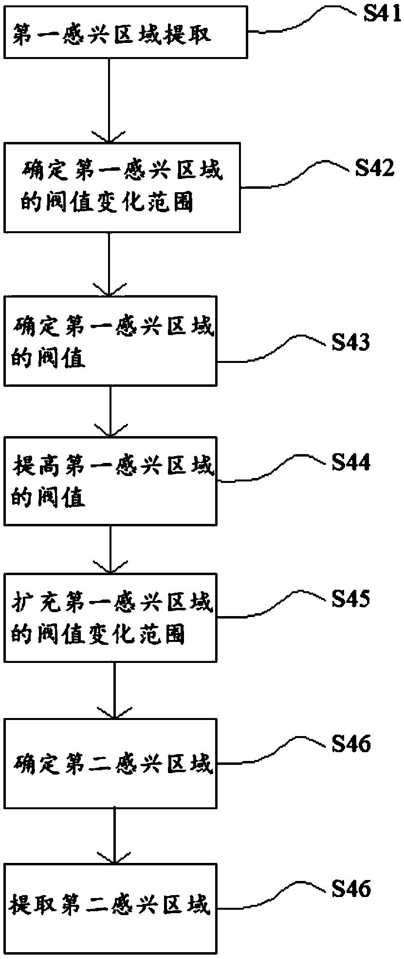
[0034] like Figure 1 ~ Figure 2 As shown, a method for segmenting pulmonary nodules based on CT images includes the following steps: Steps S1-S4. details as follows:

[0035] S1: Carry out the first binarization processing on the lung CT image data to extract the lung parenchyma region; the first binarization processing is specifically: unify the CT value for noise reduction processing, and then determine the lu...


PUM


Abstract

The invention provides a pulmonary nodule segmentation method based on a CT image. The pulmonary nodule segmentation method comprises pulmonary parenchyma region extraction, pulmonary cavity region extraction, pulmonary lobe region extraction and region of interest extraction. According to the method and the device, through the first binarization processing and the second binarization processing,the influence of the binarization processing on the extraction of the focus region, namely the first appearing region and the second appearing region, is weakened, so that the focus separation precision is improved. After the first appearing area is highlighted, the threshold value of the first appearing area is further increased, whether the coordinates marked by a doctor are accurate or not is detected, such as whether the coordinates are on the blood vessel or not, so that the accurate separation of the nodules and the blood vessel is achieved, and meanwhile the workload of manual judgmentand the workload of manual repair during later verification of the doctor are reduced. On the premise that the threshold change range of the first appearing area is reasonably expanded, the second appearing area is determined and extracted, and the focus separation precision is further improved.

Description

technical field [0001] The invention belongs to the field of medical image processing, in particular to a method for segmenting pulmonary nodules based on CT images. Background technique [0002] Lung cancer has become the malignant tumor with the highest morbidity and mortality in the world, seriously threatening human life and health. Early detection is an effective way to improve the treatment effect of lung cancer patients. The detection and identification of nodules is increasingly important in the treatment of lung cancer. Accurate segmentation of pulmonary nodules is the key content of pulmonary nodule detection and recognition research, which directly affects the reliability of pulmonary nodule-assisted diagnostic techniques. [0003] Segmenting pulmonary nodules from lung CT images is an important application of image processing technology in medical images, and is of great significance in computer-aided diagnosis. Most methods start with noise reduction and then ...

Claims


Application Information

Patent Timeline
IPC IPC(8): G06T7/11G06T7/136
CPCG06T7/11G06T7/136G06T2207/10081G06T2207/30064G06T2207/20104
Inventor 刘雷周凌霄任和
Owner 上海藤核智能科技有限公司
Why Eureka
Try Eureka