X-ray chest radiograph image quality determination method and device

An image quality and X-ray technology, applied in the field of X-ray chest film image quality determination method and device, can solve the problems affecting the safety and accuracy of diagnosis, low efficiency of manual evaluation, etc.

Active Publication Date: 2019-06-07
SHANGHAI UNITED IMAGING INTELLIGENT MEDICAL TECH CO LTD
View PDF10 Cites 16 Cited by
  • Summary
  • Abstract
  • Description
  • Claims
  • Application Information

AI Technical Summary

Problems solved by technology

[0010] 7. No extracorporeal metal and other foreign matter that X-rays cannot penetrate
[0011] Usually hospitals have a great demand for filming, but at present, the evaluation of image quality is mainly done by radiologists. Manual evaluation is inefficient, and it is easy to cause some unqualified films to enter clinical diagnosis, thus affecting the safety of diagnosis. sex and accuracy

Method used


Image

  • X-ray chest radiograph image quality determination method and device
  • X-ray chest radiograph image quality determination method and device
  • X-ray chest radiograph image quality determination method and device

Examples


Embodiment 1

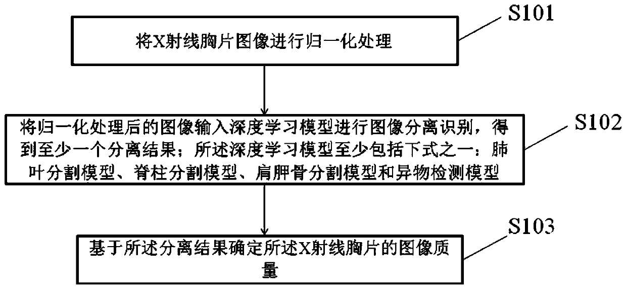
[0141] Such as figure 1 As shown, this embodiment provides a schematic flow chart of a method for determining the quality of an X-ray chest image, and this description provides the operation steps of the method as described in the embodiment or flow chart, but based on routine or non-creative work may include more more or fewer steps. The sequence of steps enumerated in the embodiments is only one of the execution sequences of many steps, and does not represent the only execution sequence. specific as figure 1 As shown, the method includes:

[0142] S101. Normalize the X-ray chest image;

[0143] S102. Input the normalized image into the deep learning model for image separation and recognition, and obtain at least one separation result; the deep learning model includes at least one of the following formulas: lung lobe segmentation model, spine segmentation model, scapula segmentation model and foreign body detection model;

[0144] The deep learning model is trained based...

Embodiment 2

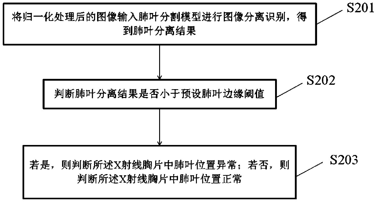
[0162] This embodiment is based on Embodiment 1. Such as image 3 As shown, when the deep learning model is a lung lobe segmentation model, the normalized image is input into the deep learning model for image separation and recognition, and at least one separation result is obtained including:

[0163] S201. Input the normalized image into the lung lobe segmentation model for image separation and recognition, and obtain the lung lobe separation result; Figure 18 As shown, where a is the image before inputting the lung lobe segmentation model; b is the output image after inputting the lung lobe segmentation model;

[0164] S202. Determine whether the lung lobe separation result is less than a preset lung lobe edge threshold;

[0165] S203. If yes, judge that the position of the lung lobe in the X-ray chest film is abnormal, and obtain the first unqualified result; if not, judge that the position of the lung lobe in the X-ray chest film is normal, and obtain the first qualifi...

Embodiment 3

[0195] Such as Figure 7 As shown, this embodiment discloses a device for determining the quality of an X-ray chest image, the device comprising:

[0196] A normalization processing module 701, configured to perform normalization processing on the X-ray chest image;

[0197] The separation result acquisition module 702 is used to input the normalized image into the deep learning model for image separation and recognition to obtain at least one separation result; the deep learning model includes at least one of the following formulas: lung lobe segmentation model, spine segmentation model , scapula segmentation model and foreign object detection model;

[0198] An image quality determining module 703, configured to determine the image quality of the X-ray chest film based on the separation result.

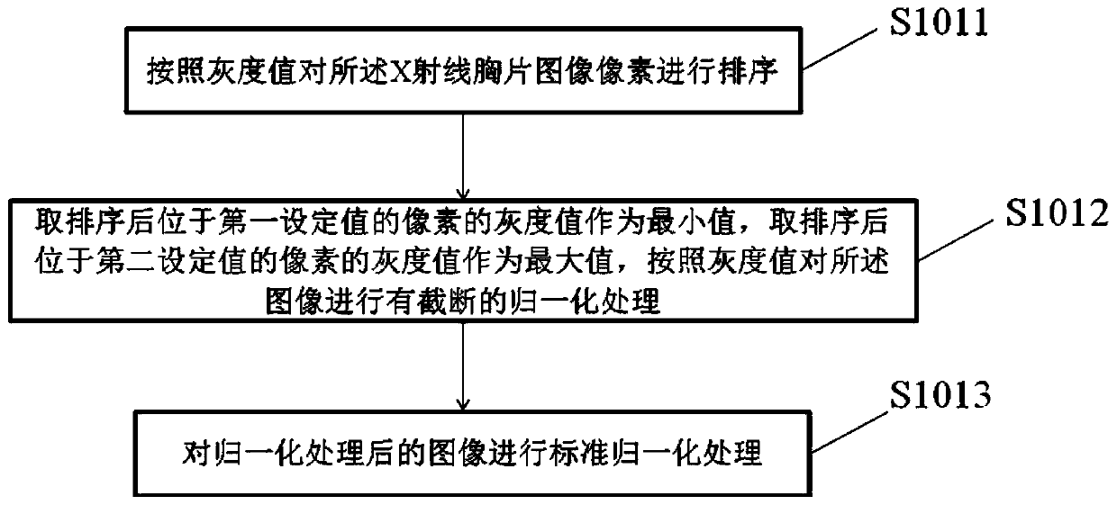
[0199] In a specific embodiment, such as Figure 8 As shown, the normalization processing module 701 includes:

[0200] A sorting unit 7011, configured to sort the X-ray chest i...


PUM


Abstract

The invention discloses an X-ray chest radiograph image quality determination method and device. The method comprises: conducting normalization processing on an X-ray chest radiograph image; inputtingthe normalized image into a deep learning model for image separation and recognition to obtain at least one separation result; wherein the deep learning model at least comprises one of the followingformulas: a pulmonary lobe segmentation model, a spine segmentation model, a shoulder blade segmentation model and a foreign matter detection model; and determining the image quality of the X-ray chest radiograph based on the separation result. The X-ray chest radiograph image quality is fully automatically evaluated, and the image quality determination speed is high. Moreover, a technician can behelped to control the image quality of the chest radiograph, and the radiograph reading accuracy is indirectly improved.

Description

technical field [0001] The invention relates to the field of X-ray chest images, in particular to a method and device for determining the quality of X-ray chest images. Background technique [0002] Chest X-ray is currently the most widely used medical image inspection method, and its usage scenarios include emergency department, intensive care, general outpatient clinic, physical examination, etc. The quality of a chest X-ray directly affects the result of diagnosis. If the quality of the chest X-ray is not up to standard, it is easy to miss and misdiagnose. [0003] Standard X-ray chest films include the following requirements: [0004] 1. The vertebral bodies below the 4th thoracic vertebra are clearly visible without bilateral shadows; [0005] 2. The texture from the hilum to the lung field is clearly displayed, and the scapula is projected on the outside of the lung field; [0006] 3. From the neck to the bifurcation of the organ, the image of the organ can be conti...

Claims


Application Information

Patent Timeline
Patent Type & Authority Applications(China)
IPC IPC(8): G06T7/00G06K9/34
CPCG06T7/0012G06T2207/20081G06T2207/30061G06T2207/30168G06T7/11G06T7/13G06T7/136
Inventor 龚再文詹恒泽郑介志詹翊强
Owner SHANGHAI UNITED IMAGING INTELLIGENT MEDICAL TECH CO LTD
Why Eureka
Try Eureka