
Method for simulating cardiac vascular lesion

A cardiovascular and vascular technology, applied in the field of cardiovascular disease simulation, can solve the problems of lack of operation experiments, difficult to perform training and teaching, and complicated operation, and achieve the goal of increasing diversity, improving authenticity, and personalized configuration and adjustment. Effect

Active Publication Date: 2019-06-07
PEKING UNION MEDICAL COLLEGE HOSPITAL CHINESE ACAD OF MEDICAL SCI +1
View PDF8 Cites 0 Cited by
  • Summary
  • Abstract
  • Description
  • Claims
  • Application Information

AI Technical Summary

Problems solved by technology

[0003] Although the above operation is a minimally invasive operation, the specific operation is very complicated due to the movement of the guide wire inside the blood vessel.
Under normal circumstances, surgical operators are required to have superb skills and rich clinical experience. However, it is difficult to implement training and teaching to improve doctors' skills. One important reason is the lack of actual operation experiments, and the patient's life cannot be used for Experimental trial and error, in this case, in order to train doctors' skills and rehearse and plan the operation process, some vascular interventional simulators specially used for simulation training came into being
[0004] In summary, although the vascular interventional simulators in the prior art can train doctors and / or medical students' surgical skills and rehearse and plan the surgical process, due to the different vascular structures and lesion locations between different patients There are differences, how to effectively improve the accuracy of the simulation process for skin coronary intervention, and the technical issues such as the authenticity of virtual training and rehearsal, are the problems that those skilled in the art need to solve

Method used


Image

  • Method for simulating cardiac vascular lesion
  • Method for simulating cardiac vascular lesion
  • Method for simulating cardiac vascular lesion

Examples


Embodiment Construction

[0038] The following examples are used to illustrate the present invention, but are not intended to limit the scope of the present invention. Unless otherwise specified, the technical means used in the embodiments are conventional means well known to those skilled in the art.

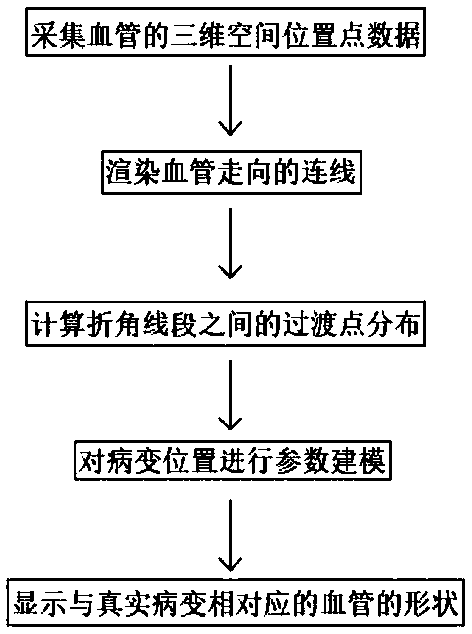
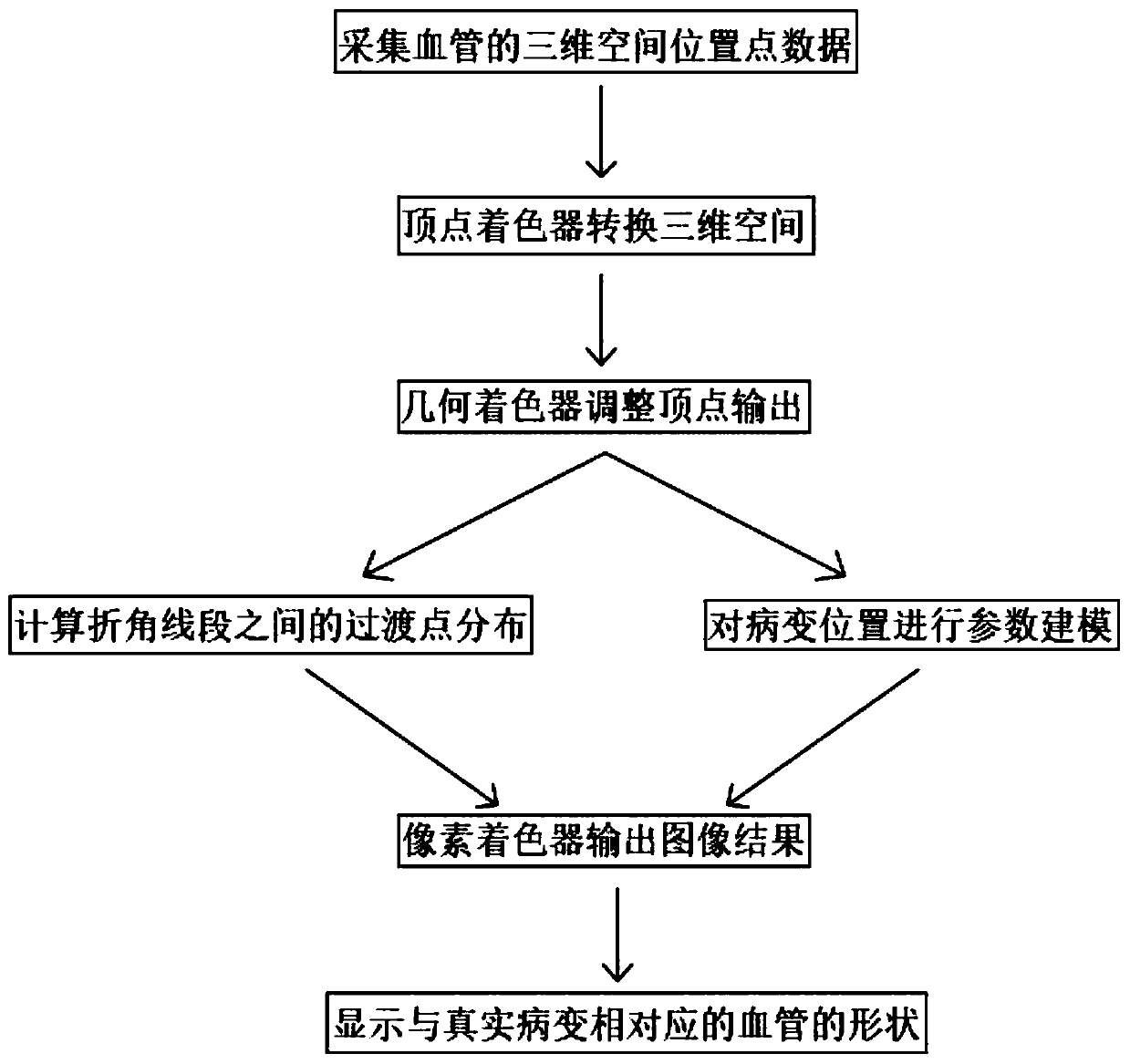
[0039] Such as figure 1 , image 3 , Figure 4 ,and Figure 5 As shown, a method for simulating cardiovascular disease includes the following steps:

[0040] Collecting the three-dimensional space position point data of blood vessels, using the connection data composed of serialized three-dimensional space data points, said three-dimensional space data points representing the blood vessel data used for simulation in three-dimensional space segmented representation;

[0041] Render the connection lines of blood vessels, and display blood vessel lines of different shapes and thicknesses based on the geometry shader;

[0042] The three-dimensional space position point data uses the serialized three-di...


PUM


Abstract

The invention discloses a method for simulating cardiac vascular lesion, and relates to the technical field of medical virtual reality. The method includes the following steps: collecting three-dimensional spatial position point data of a blood vessel; rendering a connecting line along the blood vessel, and displaying blood vessel lines with different shapes and thicknesses based on s geometric shader; calculating transition point distribution between the break angle line segments to obtain a smooth blood vessel display result; and performing parameter modeling on the lesion position, and according to the model and combined with the blood vessel display result, finally displaying the shape of the blood vessel corresponding to the real lesion. The method for simulating cardiac vascular lesion can display the true relationship between the position of the cardiac lesion and the corresponding blood vessel in a real-time environment, can more accurately simulate the actual situation of theinterventional surgery, can improve the authenticity of the doctor training and the rehearsal, and in the case of various lesions and the effects of different lesions on the deformation of blood vessels, can realize personalized configuration and adjustment and increase the diversity in the simulation process.

Description

technical field [0001] The invention relates to the technical field of medical virtual reality, in particular to a method for simulating cardiovascular disease. Background technique [0002] Cardiovascular disease is currently one of the leading causes of death. Among them, the lesions in the coronary arteries of the heart are the most serious cause of death. Usually, cardiovascular diseases mainly form plaques in the coronary arteries, and this is an important reason that seriously affects human health. An effective technique for treating cardiovascular diseases is percutaneous coronary intervention. The main process of percutaneous coronary intervention is: under the guidance of X-rays, doctors use guide wires to pass through blood vessels on the surface of the body and send them into the human thigh Artery, the guide wire passes through the femoral artery, enters the aorta retrogradely along the artery, and enters the coronary artery vascular network. Then the doctor wil...

Claims


Application Information

Patent Timeline
IPC IPC(8): G16H50/50G16H30/20
Inventor 谢洪智李帅张抒扬郝爱民王朔韩业晨杨絮飞
Owner PEKING UNION MEDICAL COLLEGE HOSPITAL CHINESE ACAD OF MEDICAL SCI
Learn More