Liver segmentation method based on three-dimensional image segmentation algorithm

A technology of three-dimensional image and liver, which is applied in the field of medical image segmentation and processing, can solve the problems of manual setting of large parameters, large influence of the initial value of SVM algorithm, and noise sensitivity, so as to avoid the influence of algorithm robustness, accurate and automatic liver Segmentation, the effect of a high level of automation

Active Publication Date: 2019-06-25
安徽紫薇帝星数字科技有限公司
View PDF3 Cites 5 Cited by
  • Summary
  • Abstract
  • Description
  • Claims
  • Application Information

AI Technical Summary

Problems solved by technology

[0004] 1. Morphological segmentation methods. For example, Lim uses multi-threshold combined with morphological filtering to extract the initial contour of the liver, and uses the gradient information and gray distribution information near the contour to obtain the final result. The disadvantage of this method is that it needs to manually set a large number of parameters. The accuracy of the segmentation results has a great influence, and this method is only suitable for images with a large grayscale difference between the liver and surrounding organs;
[0005] 2. Segmentation methods based on deformation models, such as Heimann combining deformation models and statistical priors for segmentation, but segmentation methods based on deformation models need to use a large number of liver shape pictures to train the statistical shape model to obtain the outline of the liver. This method takes a long time and the segmentation results are greatly affected by the training pictures;
[0006] 3. Neural network-based segmentation methods, such as Wang improved the fuzzy cellular neural network and applied it to liver segmentation, Zafer proposed a new supervised learning neural network ISNN (incremental supervised neural network), and applied it to Liver segmentation, etc. This method needs to artificially establish a template from the segmentation result, and the segmentation result is greatly affected by the template;
[0007] 4. Based on clustering segmentation methods, such as Liu combined K-means and SVM for liver segmentation, but the SVM algorithm is greatly affected by the initial value, and is sensitive to noise, and the stability of the algorithm is low

Method used


Image

  • Liver segmentation method based on three-dimensional image segmentation algorithm
  • Liver segmentation method based on three-dimensional image segmentation algorithm
  • Liver segmentation method based on three-dimensional image segmentation algorithm

Examples


Embodiment Construction

[0032] The following will clearly and completely describe the technical solutions in the embodiments of the present invention with reference to the accompanying drawings in the embodiments of the present invention. Obviously, the described embodiments are only some, not all, embodiments of the present invention. All other embodiments obtained by persons of ordinary skill in the art based on the embodiments of the present invention belong to the protection scope of the present invention.

[0033] According to an embodiment of the present invention, a liver segmentation method based on a three-dimensional graph cut algorithm is provided.

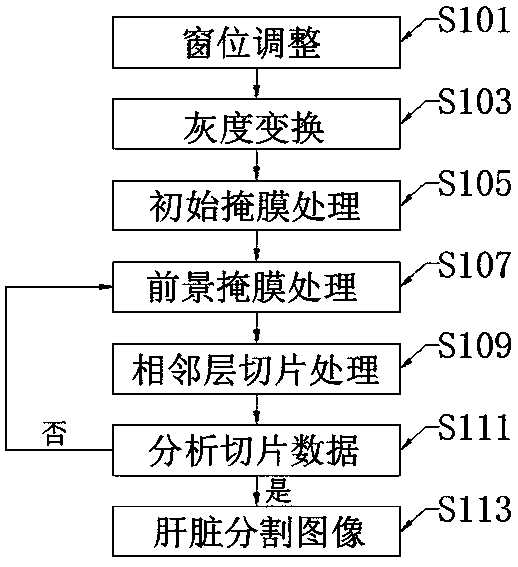
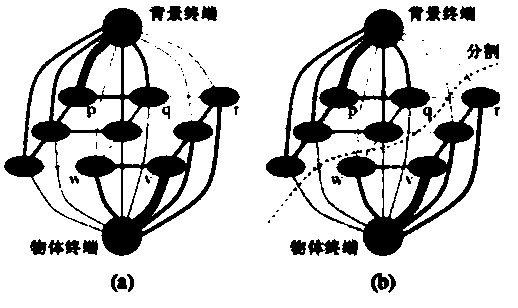
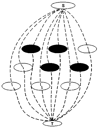
[0034] Such as Figure 1-3 As shown, the liver segmentation method based on the three-dimensional graph cut algorithm according to the embodiment of the present invention includes the following steps:

[0035] S101. Window level adjustment: adjust the window width and window level of the CT image sequence in advance to highlight the developme...


PUM


Abstract

The invention discloses a liver segmentation method based on a three-dimensional image segmentation algorithm, and the method comprises the following steps: S101, carrying out the window position adjustment: carrying out the adjustment of the window width and the window position of a CT image sequence in advance, highlighting the development of a liver region, and obtaining an adjustment image CTimage A; S103, gray scale transformation: carrying out gray scale transformation processing on the obtained CT image A of the adjustment image, keeping a liver region image, and filtering out a dark tissue image to obtain an enhanced image CT image B; and S105, performing initial mask processing: randomly selecting a single slice in the abdominal CT image from the obtained enhanced image B, and performing liver two-dimensional segmentation on the single slice in the abdominal CT image sequence by using a GraphCut algorithm. According to the method, the liver region of the CT image is segmentedthrough the three-dimensional image segmentation algorithm, liver segmentation can be rapidly completed through iteration according to a single liver segmentation result in the three-dimensional CT image, complete liver image information is obtained, subsequent reconstruction is facilitated, and rapid, accurate and automatic liver segmentation can be achieved.

Description

technical field [0001] The invention relates to the technical field of medical image segmentation processing, in particular to a liver segmentation method based on a three-dimensional graph cut algorithm. Background technique [0002] Liver cancer is the most common malignant liver disease with a high mortality rate. Computed tomography (CT, Computer Tomography), as a non-invasive and accurate imaging method for the diagnosis of liver cancer, not only enables doctors to obtain important data and information of lesions more directly and clearly, but also saves patients from the pain of invasive diagnosis, but a large number of The analysis of CT images is time-consuming and may lead doctors to make wrong judgments. Therefore, the study of Computer Aided Diagnosis (CAD) system is of great significance and value to help doctors improve diagnosis efficiency. As one of the most critical steps in the liver computer-aided diagnosis system, the segmentation of liver CT images is t...

Claims


Application Information

Patent Timeline
Patent Type & Authority Applications(China)
IPC IPC(8): G06T7/11G06T7/194
Inventor 王宜主欧阳挺仲红艳居庆玮
Owner 安徽紫薇帝星数字科技有限公司
Why Eureka
Try Eureka