
Liver tumor segmentation method and device based on deep learning

A deep learning technology for liver tumors, applied in image analysis, image data processing, instruments, etc., can solve the problems of not considering the spatial structure information of three-dimensional data tumors, mining, and inaccurate segmentation results, and achieve high tumor segmentation accuracy and improve Robustness, the effect of improving segmentation accuracy

Inactive Publication Date: 2019-06-25
BEIJING INSTITUTE OF TECHNOLOGYGY
View PDF2 Cites 24 Cited by
  • Summary
  • Abstract
  • Description
  • Claims
  • Application Information

AI Technical Summary

Problems solved by technology

However, most neural network-based methods only use the two-dimensional slice information of the tumor without considering the spatial structure information contained in the three-dimensional slice data, which leads to the lack of full use of the continuity of the tumor in the spatial structure during model training. information resulting in inaccurate segmentation results
[0004] Although the research on automatic segmentation of liver tumors has received extensive attention and achieved certain research results, there are still the following problems: the size, shape, location, and number of tumors in different patients with liver tumors are different, making the segmentation based on domain prior knowledge The effect of the tumor segmentation method is not good; because the edge of the liver tumor is not clear, the manual feature extraction method can not make the model learn the difference between tumor and non-tumor pixels, so most of the discriminative segmentation methods based on pixel intensity information are in There are still some difficulties in segmenting tumor margins; most of the segmentation methods based on deep learning only use two-dimensional slice information without considering the tumor spatial structure information contained in three-dimensional data, and a few other methods that use three-dimensional data for network training are affected by Due to the limitation of computing resources, only a very small number of slices are used, which makes it difficult to effectively mine the spatial structure of the tumor, making it difficult for the model to capture the global characteristics of the tumor in the 3D image sequence, and lose the unique features of the tumor in the sequence image. Spatial continuity information leads to unsatisfactory segmentation results

Method used


Image

  • Liver tumor segmentation method and device based on deep learning
  • Liver tumor segmentation method and device based on deep learning
  • Liver tumor segmentation method and device based on deep learning

Examples

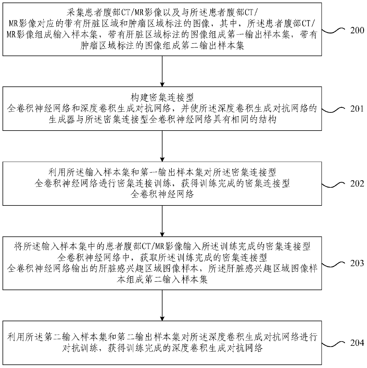

Embodiment Construction

[0025] In order to make the purpose, technical solutions and advantages of the embodiments of the present invention clearer, the technical solutions in the embodiments of the present invention will be clearly and completely described below in conjunction with the drawings in the embodiments of the present invention. Obviously, the described embodiments It is a part of embodiments of the present invention, but not all embodiments. Based on the embodiments of the present invention, all other embodiments obtained by persons of ordinary skill in the art without creative efforts fall within the protection scope of the present invention.

[0026] The embodiment of the present invention combines the fully convolutional neural network and generative adversarial network technology in deep learning, and cooperates with the liver gold standard and tumor gold standard manually drawn by the doctor as the input of the network for learning, so that the network can automatically capture the li...


PUM


Abstract

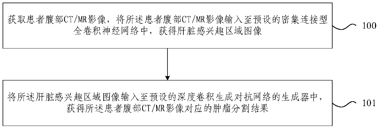
The embodiment of the invention provides a liver tumor segmentation method and device based on deep learning, and the method comprises the steps: obtaining an abdominal CT / MR image of a patient, inputting the abdominal CT / MR image of the patient into a preset dense connection type full convolutional neural network, and obtaining a liver region-of-interest image; and inputting the liver region-of-interest image into a preset deep convolutional generative adversarial network generator to obtain a tumor segmentation result. According to the embodiment of the invention, through adoption of the densely connected full convolutional neural network and the deep convolutional generative adversarial network, the robustness and segmentation accuracy of liver tumor segmentation are improved, and the features extracted in the two-dimensional plane and the structure features extracted in the three-dimensional space are fused, so that the tumor segmentation precision is higher.

Description

technical field [0001] Embodiments of the present invention relate to the field of liver tumor segmentation based on CT / MR imaging, and more specifically, relate to a method and device for liver tumor segmentation based on deep learning. Background technique [0002] Using computer algorithms to realize automatic segmentation of liver tumors in abdominal CT / MR slices can provide accurate and repeatable tumor detection services, assist doctors in diagnosis, and play an important role in surgical planning and tumor treatment evaluation. Research hotspots in the field of medical image processing. However, liver tumor segmentation has always been a big challenge, because the size, shape, and location of tumors vary greatly among different patients, which limits many methods for tumor segmentation based on prior information such as shape and location. In addition, due to the unclear boundary between the liver tumor and its surrounding normal tissue, many traditional segmentation...

Claims


Application Information

Patent Timeline
IPC IPC(8): G06T7/11G06N3/04
Inventor 宋红陈磊杨健艾丹妮
Owner BEIJING INSTITUTE OF TECHNOLOGYGY
Learn More