X-ray two-dimensional phase-contrast imaging method of single exposure

A phase-contrast imaging and X-ray technology, applied in the field of X-ray imaging, can solve the problems of reducing the ability of defect detection and detail resolution, complicated acquisition process, and increased imaging time, so as to improve the ability of object detail resolution and defect detection, simplify Collection process, effect of radiation dose reduction

Active Publication Date: 2019-07-05
LANZHOU UNIVERSITY
View PDF7 Cites 9 Cited by
  • Summary
  • Abstract
  • Description
  • Claims
  • Application Information

AI Technical Summary

Problems solved by technology

[0006] However, at present, the edge illumination method needs to move the coded diaphragm once, perform an exposure at two positions respectively, and collect two original images to quantitatively extract phase and absorption information, resulting in complicated acquisition process and increased imaging time, which is not conducive to practical application
In addition, the edge illumination method only realizes one-dimensional phase contrast imaging. Since the phase information ...

Method used


Image

  • X-ray two-dimensional phase-contrast imaging method of single exposure
  • X-ray two-dimensional phase-contrast imaging method of single exposure
  • X-ray two-dimensional phase-contrast imaging method of single exposure

Examples


Embodiment 1

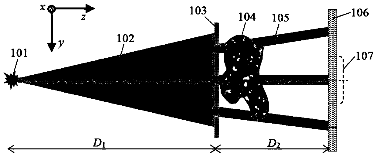
[0038] see Figure 1-7 As shown, a single-exposure X-ray two-dimensional phase-contrast imaging method, its structure includes an X-ray source 101, a coding aperture M-103 and an area array detector 106, and the method specifically includes the following steps:

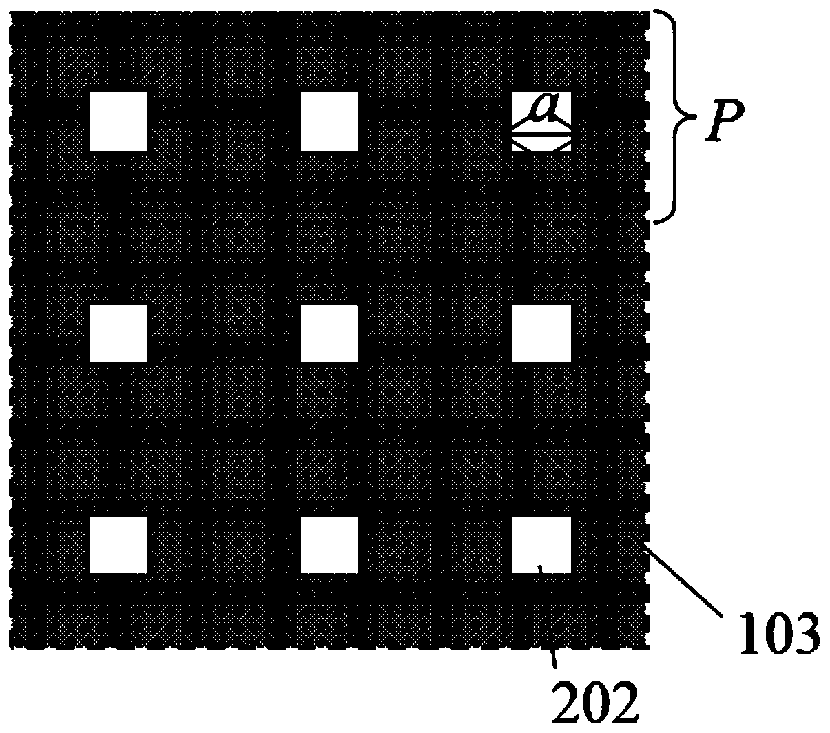
[0039]Step 1: The coded diaphragm M-103 is placed in front of the X-ray source 101, and the X-ray beam is periodically divided into many thin beams. The period P of the coded diaphragm M-103 is determined by the pixel size d of the area array detector 106, And the distance D from the X-ray source 101 to the coding aperture M-103 1 And the distance D from the encoding aperture M-103 to the area array detector 106 2 jointly determined, can be expressed as P=2d / m, where m is the amplification factor, can be expressed as m=(D 1 +D 2 ) / D 1 , the opening size of the coded diaphragm M-103 is a;

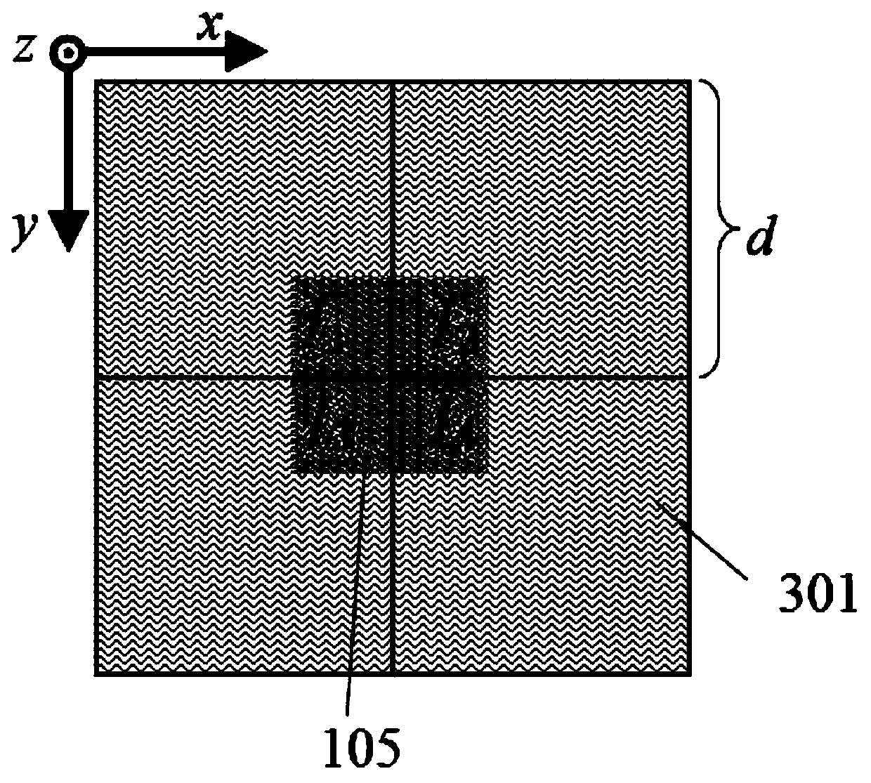
[0040] Step 2: using four adjacent pixels of the area array detector 106 as a detection unit 107, the X-ray sub-beam 105 emit...

Embodiment 2

[0059] Due to the influence of pixel crosstalk, image contrast and line-to-noise ratio will be reduced. In this embodiment, coding aperture M2 408 is added to block adjacent areas of pixels to reduce the influence of pixel crosstalk, such as Figure 4 shown. In order to compare with the aforementioned imaging method, the experimental conditions of Example 2 are the same as those of Example 1 except that the coding aperture M-408 is added and the opening width a of the coding aperture M-103 is set to 32 μm. The case where the coding aperture M2408 blocks the detector unit and the X-ray irradiation conditions are as follows Figure 5 As shown, the coding aperture M2 408 blocks the width b of the edge of the pixel to 20 μm. This condition makes the X-ray irradiation area of ​​the four pixels in the second embodiment the same as that in the first embodiment. Carry out numerical simulation experiments based on the above experimental conditions, and use the imaging method proposed ...


PUM

PropertyMeasurementUnit
Thicknessaaaaaaaaaa
Thicknessaaaaaaaaaa
Opening widthaaaaaaaaaa

Abstract

The invention discloses an X-ray two-dimensional phase-contrast imaging method of single exposure. The method specifically includes following steps: during imaging, a detected object is arranged between a first encoding diaphragm M and an area array detector and close to the first encoding diaphragm M, the change of the intensity and the direction of the X-ray is caused by absorption and refraction of the X-ray by the detected object, the ray intensities I1, I2, I3 and I4 detected by pixels of the detector are changed, quantified phase and absorption information can be extracted by employing relative changes thereof, and two-dimensional phase-contrast imaging of the X-ray of single exposure is realized. The beneficial effects of the method are that the acquisition process is simplified, original secondary exposure is simplified as single exposure, the imaging time and the irradiation dose are reduced by 50% compared with that of the original method, the mode of single exposure can realize dynamic and online phase-contrast imaging, the realization of the phase-contrast CT technology of the X-ray is facilitated, two-dimensional phase-contrast imaging can be realized, and the object detail distinguishing capability and the defect detection capability are improved.

Description

technical field [0001] The invention belongs to the field of X-ray imaging, in particular to an X-ray two-dimensional phase-contrast imaging method that only needs a single exposure. Background technique [0002] In traditional X-ray imaging, the image contrast is based on the object’s absorption of X-rays. For strongly absorbing objects, it can give a higher image contrast, but for weakly absorbing objects, such as human soft tissue and fiber materials, the image contrast is low. In addition, for objects with similar absorption characteristics, the density resolution ability is limited, and it is difficult to accurately distinguish different materials. For example, in the early diagnosis of tumors, the detection sensitivity is low due to the close density of diseased tissue and normal tissue. X-ray Phase-Contrast Imaging (XPCI) can solve the above problems, giving higher image contrast and clearer object details. [0003] As an electromagnetic wave, X-rays will not only at...

Claims


Application Information

Patent Timeline
IPC IPC(8): G01N23/041G01N23/046
CPCG01N23/041G01N23/046G01N2223/03G01N2223/1016
Inventor 张催潘小东李公平商宏杰
Owner LANZHOU UNIVERSITY
Why Eureka
Try Eureka