FISH probe set for detecting FGFR2 fusion and application thereof
A probe group and probe technology, applied in the direction of recombinant DNA technology, microbial measurement/testing, DNA/RNA fragments, etc., to achieve high specificity and sensitivity, reduce drug side effects, and ingeniously designed effects
- Summary
- Abstract
- Description
- Claims
- Application Information
AI Technical Summary
Problems solved by technology
Method used
Image
Examples
Embodiment 1
[0030] The FGFR2 5' end probe set used two BAC clones (RP11-78A18 and RP11-436E17) as templates, and the 3' end probe set used two BAC clones (RP11-7P17 and RP11-984I17) as templates, and the above BAC clones were all Purchased from Oakland Children's Hospital Research Institute, USA. The fabrication and labeling steps of the probe set are as follows:
[0031] (1) In two 200 μL PCR reaction tubes, add the reaction materials according to the following system to prepare and label the 5′-end probe set and the 3’-end probe set:
[0032]
[0033]
[0034] The above random primers consist of 9 nucleotides, each nucleotide is A, T, C or G, and the first and / or fifth nucleotides from the 5' end are modified with fluorescence through spacer9 connection group; the formula of the above 5×Buffer is: 10mM Tris-HCl, 25mM MgCl 2 (magnesium chloride), with ddH 2 O preparation.
[0035] After mixing by pipetting, denature at 95°C for 10 minutes on a PCR instrument, immediately place ...
Embodiment 2
[0049] The steps of using a UV spectrophotometer to measure the labeling efficiency and concentration of the probes in the above-mentioned probe group are as follows:
[0050] (1) Turn on the UV spectrophotometer (NanoDrop), select the full-wavelength mode, and select the corresponding wavelength. After blank calibration, measure the A of the 5′-end probe group respectively. 260nm 、A Dye (FITC:A 494nm ) and A of the 3′ end probe set 260nm 、A Dyc (Tetramethyl-rhodamine: A 560nm ).
[0051] (2) Since the fluorescent dye also has a certain absorbance value at 260nm, in order to make the absorbance value of the probe at 260nm more accurate, use the following formula to correct A 260 Value: A Base =A 260 -(A Dye ×CF 260 ), CF 260 Refers to the correction factor, which is a constant, and each fluorescent dye has a certain CF 260 Values, such as the CF of Tetramethyl-rhodamine 260 The value is 0.06 and FITC is 0.32. (3) Calculate the probe labeling efficiency and concent...
Embodiment 3
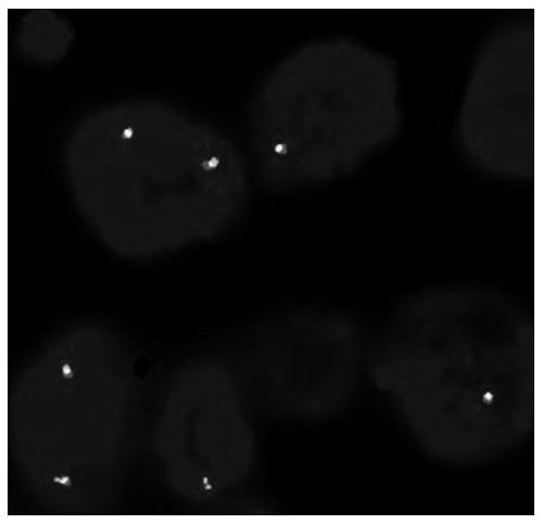
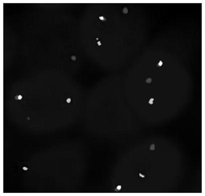
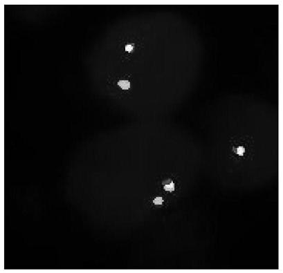
[0056] The FGFR2 gene breakage probe system prepared in Example 1 was used to detect 50 intrahepatic cholangiocarcinoma samples with known FGFR2 gene fusion results provided by the hospital, and the detection steps were as follows:
[0057] (1) Sample dewaxing and rehydration: immerse tissue section samples in 3 tanks of xylene for 10 minutes each time for dewaxing, and then soak in 100%, 100%, 100%, 85% and 70% ethanol for 3 minutes, Finally, immerse in deionized water for 3 min.
[0058] (2) Sample pretreatment and digestion: Boil the tissue section sample in 100°C water for 20 minutes, take out the sample and digest it with pepsin for 10 minutes, transfer it to 2×SSC solution for 3 minutes, take out the sample and immerse it in 70%, 85% and 100% Ethanol dehydration, 3min each time, take out the sample to dry naturally.
[0059] (3) Hybridization of the sample with the probe: Use a pipette gun to take 10 μL of the FGFR2 gene fragmentation probe system and add it to the samp...
PUM
 Abstract
Description
Claims
Application Information
 - R&D
- Intellectual Property
- Life Sciences
- Materials
- Tech Scout
- Unparalleled Data Quality
- Higher Quality Content
- 60% Fewer Hallucinations
Browse by: Latest US Patents, China's latest patents, Technical Efficacy Thesaurus, Application Domain, Technology Topic, Popular Technical Reports.
© 2025 PatSnap. All rights reserved.Legal|Privacy policy|Modern Slavery Act Transparency Statement|Sitemap|About US| Contact US: help@patsnap.com


