Medical image examination equipment for cardiovascular medicine department

A technology of internal medicine and inspection equipment, applied in medical science, patient positioning for diagnosis, instruments for radiological diagnosis, etc., can solve problems such as inconvenience, reduce labor, and ensure the effect of shooting quality

Active Publication Date: 2019-07-12
FUWAI HOSPITAL CHINESE ACADEMY OF MEDICAL SCI
View PDF13 Cites 6 Cited by
  • Summary
  • Abstract
  • Description
  • Claims
  • Application Information

AI Technical Summary

Problems solved by technology

At present, when patients undergo heart imaging examinations, the general patients give way to standing positions, and patients need to constantly change an

Method used


Image

  • Medical image examination equipment for cardiovascular medicine department
  • Medical image examination equipment for cardiovascular medicine department
  • Medical image examination equipment for cardiovascular medicine department

Examples


Example Embodiment

[0029] A specific embodiment of the present invention will be described in detail below with reference to the accompanying drawings, but it should be understood that the protection scope of the present invention is not limited by the specific embodiments.

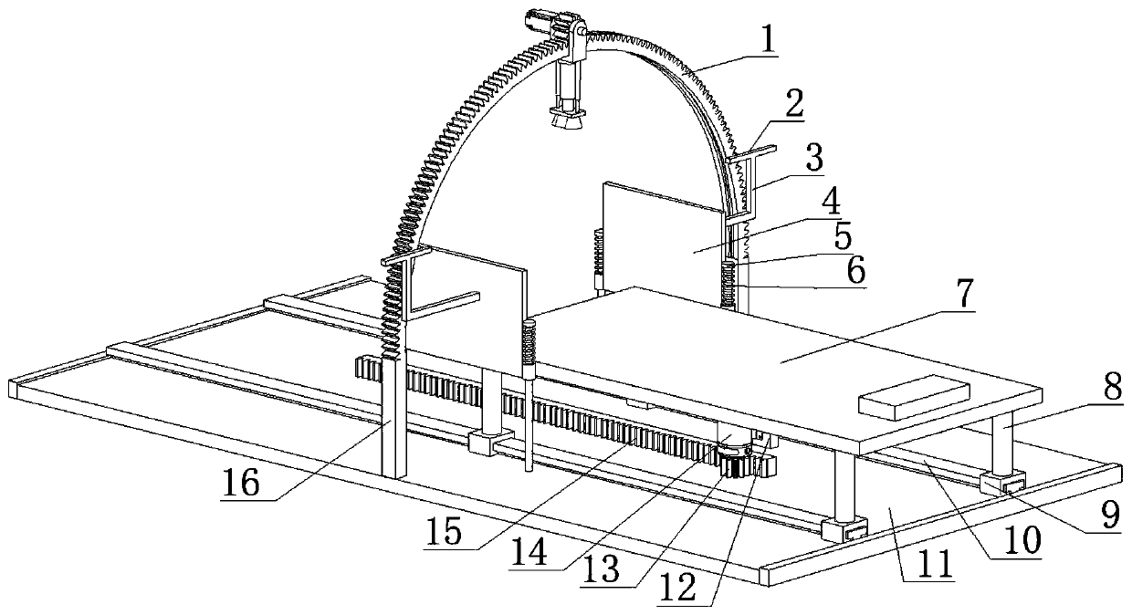
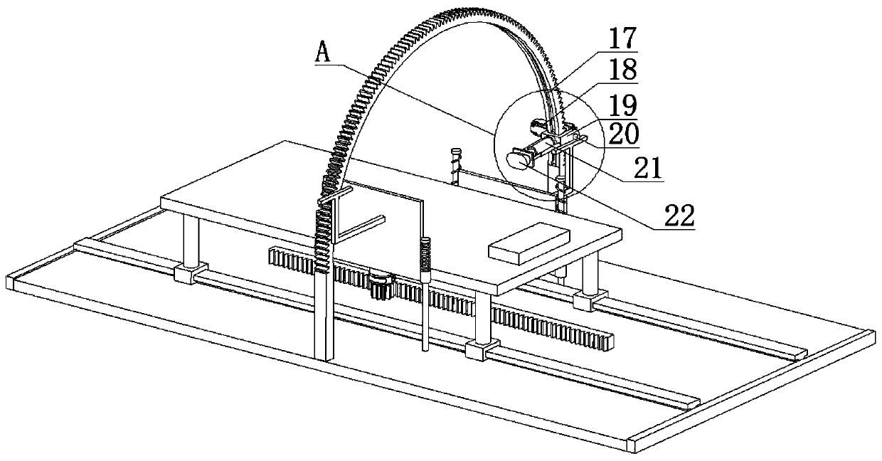
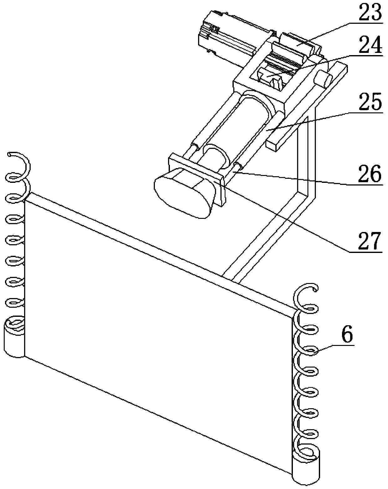
[0030] Such as Figure 1-Figure 11 As shown, the present invention includes a bottom plate 11, the two sides of the upper middle of the bottom plate 11 are respectively fixedly connected with support columns 16, the upper ends of the two support columns 16 are respectively fixedly connected to the two ends of the half gear ring 1, the half tooth The inner side of the ring 1 is provided with a T-shaped arc-shaped sliding groove 17 respectively. The T-shaped arc-shaped sliding groove 17 is provided with a T-shaped arc-shaped sliding block 24, and the outer end of the T-shaped arc-shaped sliding block 24 is fixedly connected to In the middle of the inner side of the U-shaped plate 19, the U-shaped plate 19 is provided with a gear...


PUM


Abstract

The invention relates to the field of image examination, in particular to medical image examination equipment for the cardiovascular medicine department. The equipment comprises a bottom plate and ischaracterized in that supporting columns are respectively and fixedly connected with the two sides of the middle of the upper side of the bottom plate, the upper ends of the two supporting columns arerespectively and fixedly connected with the two ends of a semi-gear ring, and the inner side of the semi-gear ring is provided with a T-shaped arc slide groove; a T-shaped arc slide block is arrangedin the T-shaped arc slide groove, the external end of the T-shaped arc slide block is fixedly connected with the middle of the inner side of a U-shaped plate, and a first gear is arranged in the U-shaped plate. According to the equipment, a patient lies in a bed, it is not necessary to require the patient to constantly change and adjust body positions, for a patient with a mobility problem, the examination is quite convenient, and the labor amount of nurses is reduced.

Description

technical field [0001] The invention relates to the field of image inspection, in particular to a medical image inspection device for cardiovascular medicine. Background technique [0002] Cardiac X-ray plain film is one of the important basis for doctors to diagnose and treat patients with heart disease. The treatment effect can be seen by comparing the X-ray plain film of the patient's heart size before and after treatment. Cardiovascular conventional chest X-ray examinations include anteroposterior view (focal-film distance 200cm), left anterior oblique view (60°-65°), right anterior oblique view (45°-55°), left and right views. Frontal chest radiography can show the size, shape, position and outline of the great vessels of the heart, and can observe the relationship between the heart and adjacent organs and the changes of blood vessels in the lungs, and can be used to measure the heart and its diameter. The left anterior oblique view shows the general picture of the ao...

Claims


Application Information

Patent Timeline
IPC IPC(8): A61B6/00A61B6/04
CPCA61B6/0407A61B6/44A61B6/503
Inventor 刘曦华伟
Owner FUWAI HOSPITAL CHINESE ACADEMY OF MEDICAL SCI
Why Eureka
Try Eureka