Tissue slice classification method based on microscopic hyperspectral imaging technology

A technology of hyperspectral imaging and tissue sectioning, applied in the field of medical image signal processing, can solve the problems that algorithms and simple discriminant models are difficult to extract and discriminate classification, image features are not obvious enough, and cannot meet the requirements of precise medical disease accurate positioning, etc. Achieve the effect of perfecting the automatic data collection and classification process, and improving the classification accuracy and speed

Active Publication Date: 2019-07-19
西安康汇馨光检科技有限公司
View PDF6 Cites 10 Cited by
  • Summary
  • Abstract
  • Description
  • Claims
  • Application Information

AI Technical Summary

Problems solved by technology

The results do not meet the requirements of precision medicine for accurate positioning of diseases, especially in the face of more complex disease subtypes and branches, the image features are not obvious enough, and the difference in spectral features is small, which is difficult to extract by traditional algorithms and simple discriminant models Deep features for efficient discriminative classification

Method used


Image

  • Tissue slice classification method based on microscopic hyperspectral imaging technology
  • Tissue slice classification method based on microscopic hyperspectral imaging technology
  • Tissue slice classification method based on microscopic hyperspectral imaging technology

Examples


Embodiment Construction

[0046] In order to make the technical solutions and advantages of the present invention clearer, the present invention will be described in more detail below in conjunction with the accompanying drawings and specific embodiments.

[0047] Such as figure 1 As shown, the microscopic hyperspectral imager is composed of a hyperspectral imaging system, a biological microscope system and a control computer. The system contains 256 bands in total, the spectral range is 400nm-1000nm, the average spectral resolution is 3nm, and the spatial resolution can reach 0.5μm. The image size is 753*696.

[0048] Microscopic objectives with different magnifications (for example: 4×, 10×, 20×, 40×, 100×) can be selected according to different objectives during the experiment, adjust the intensity of the light source, pay attention not to saturate, and adjust the focusing mechanism to ensure that the sample is in the best condition position, select the target area, and collect microscopic hyperspe...


PUM

PropertyMeasurementUnit
Sensitivityaaaaaaaaaa

Abstract

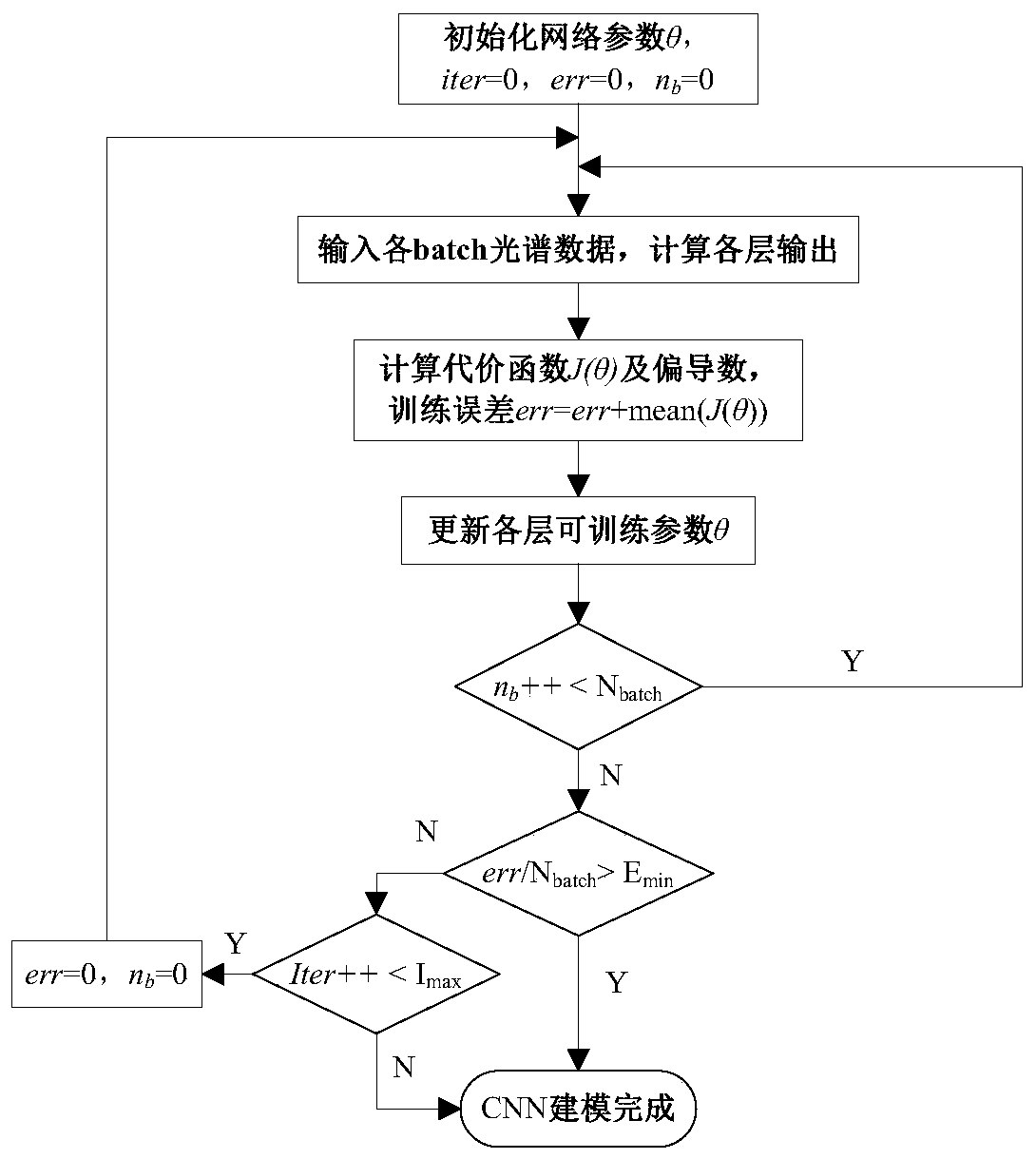
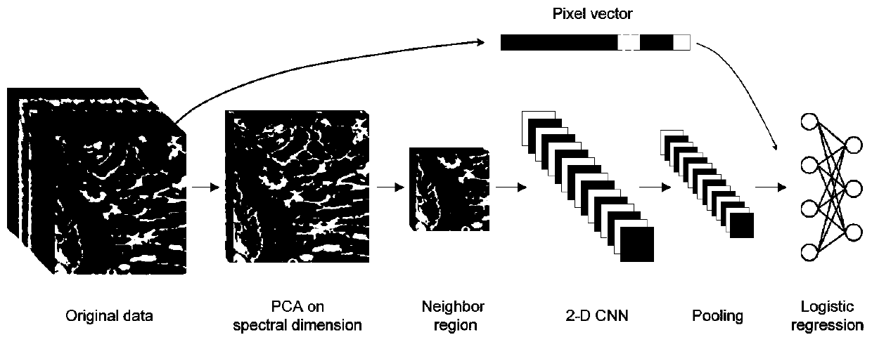
The invention provides a tissue slice classification method based on a microscopic hyperspectral imaging technology. The method comprises the following steps of firstly, preprocessing the microscopichyperspectral data so as to eliminate the noise influence and the data redundancy; establishing and training three types of convolutional neural network (CNN) models; wherein the one-dimensional CNN model realizes the spectral dimension feature extraction and classification, and the two-dimensional CNN model and the three-dimensional CNN model realize the spectrum-spectral dimension joint featureextraction and classification respectively; and for an actual to-be-detected microscopic hyperspectral image, obtaining a final classification result through the quantitative qualitative analysis andthe model output result voting. According to the present invention, the deep learning convolutional neural network models are used for extracting and classifying the deep features, so that the overallclassification precision and the speed are improved, and meanwhile the automatic data collection and classification process of the pathological sections is perfected.

Description

technical field [0001] The invention belongs to the technical field of medical image signal processing, and in particular relates to a method for classifying tissue slices based on microscopic hyperspectral imaging technology. Background technique [0002] Medical hyperspectral imaging is a comprehensive cross-cutting technology based on clinical medicine, imaging, medical sensors, pathological tissue analysis and other technologies. It belongs to the new field application of hyperspectral technology in recent years. [0003] The microscopic hyperspectral technology integrated with the microscopic imaging system can obtain microscopic hyperspectral images with higher spatial resolution, making it possible to study microscopic objects (such as tissue slices, cells, microorganisms, etc.). Microscopic hyperspectral imaging technology combines two traditional optical diagnostic technologies of spectral analysis and optical imaging, and can simultaneously provide information on t...

Claims


Application Information

Patent Timeline
Patent Type & Authority Applications(China)
IPC IPC(8): G06K9/62G01N21/25G06N3/04G06N3/08
CPCG06N3/08G01N21/25G06N3/044G06N3/045G06F18/2411G06F18/254
Inventor 胡炳樑杜剑张周锋于涛
Owner 西安康汇馨光检科技有限公司
Why Eureka
Try Eureka