
Image processing method and device and storage medium

An image processing and image technology, applied in the field of image processing, to achieve the effect of improving efficiency, improving flexibility, and increasing speed

Active Publication Date: 2019-08-16
SHANGHAI SENSETIME INTELLIGENT TECH CO LTD
View PDF6 Cites 9 Cited by
  • Summary
  • Abstract
  • Description
  • Claims
  • Application Information

AI Technical Summary

Problems solved by technology

Currently, there is no image processing method for accurate lesion boundary detection.

Method used


Image

  • Image processing method and device and storage medium
  • Image processing method and device and storage medium
  • Image processing method and device and storage medium

Examples


Embodiment Construction

[0053] The following will clearly and completely describe the technical solutions in the embodiments of the application with reference to the drawings in the embodiments of the application. Apparently, the described embodiments are only some of the embodiments of the application, not all of them. Based on the embodiments in this application, all other embodiments obtained by persons of ordinary skill in the art without making creative efforts fall within the protection scope of the present invention.

[0054] This application is mainly applied to image processing equipment, which can be traditional image processing equipment, computer-aided diagnosis equipment, remote diagnosis equipment, pathological image labeling equipment, online teaching equipment of medical schools, or the third and fourth embodiments of this application. The image processing device described in the embodiment is not limited in this application. When the image processing device interacts with other devic...


PUM


Abstract

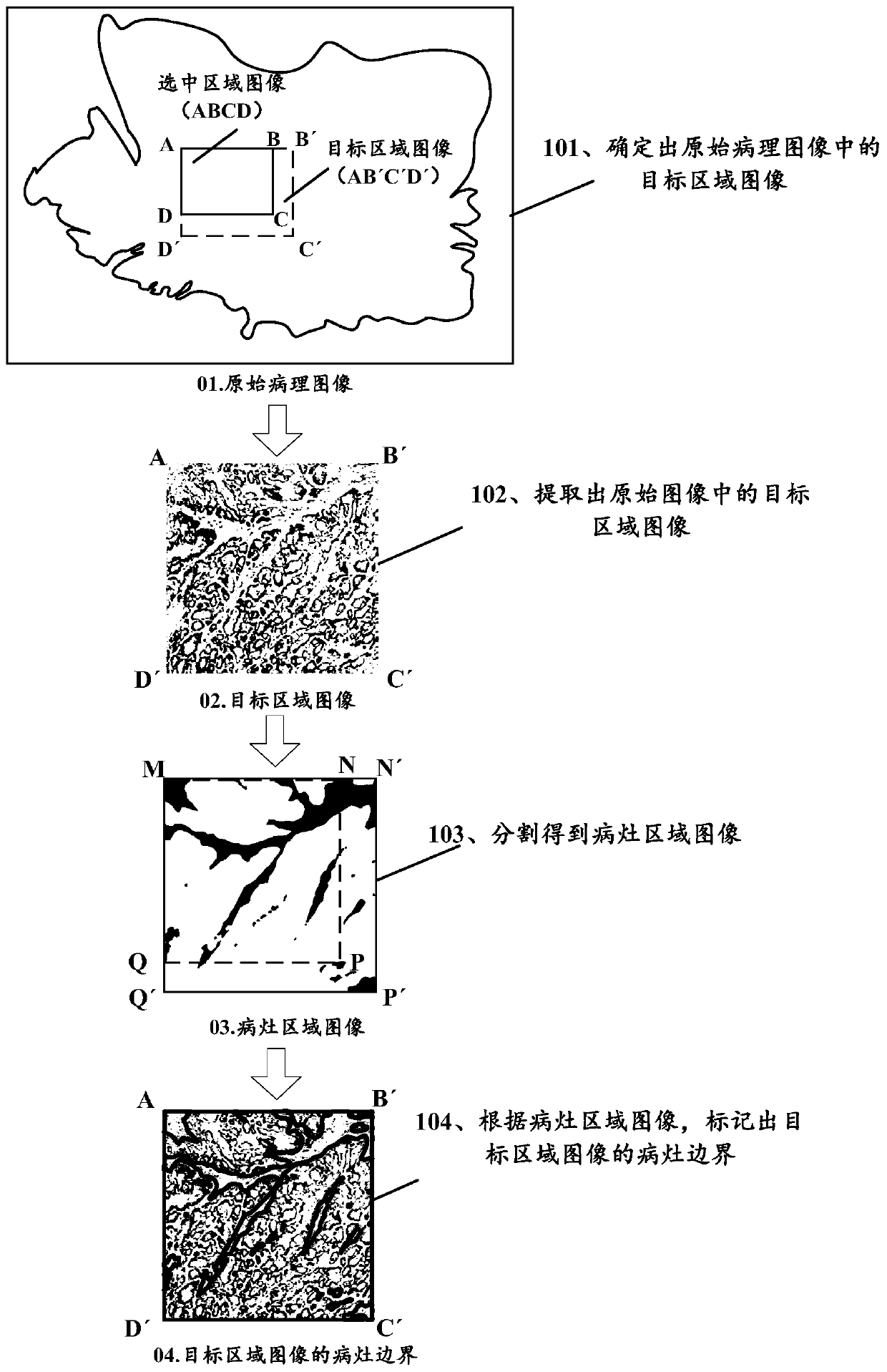
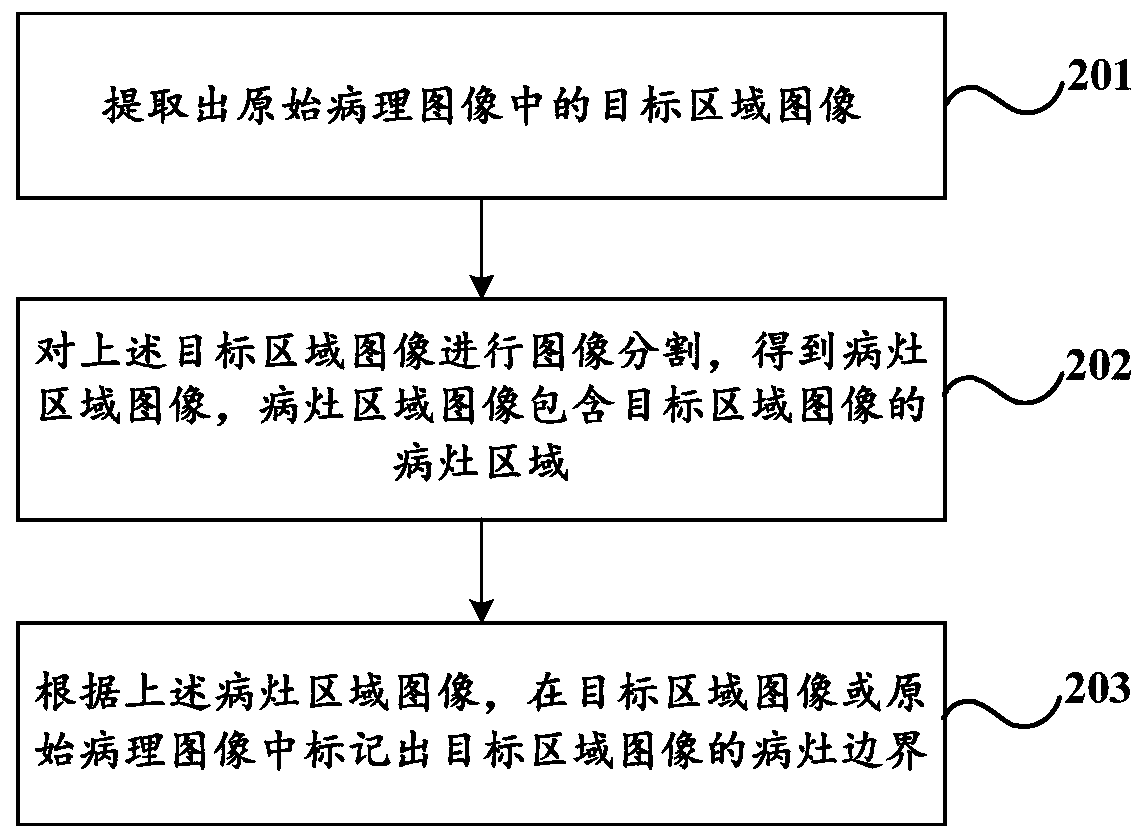
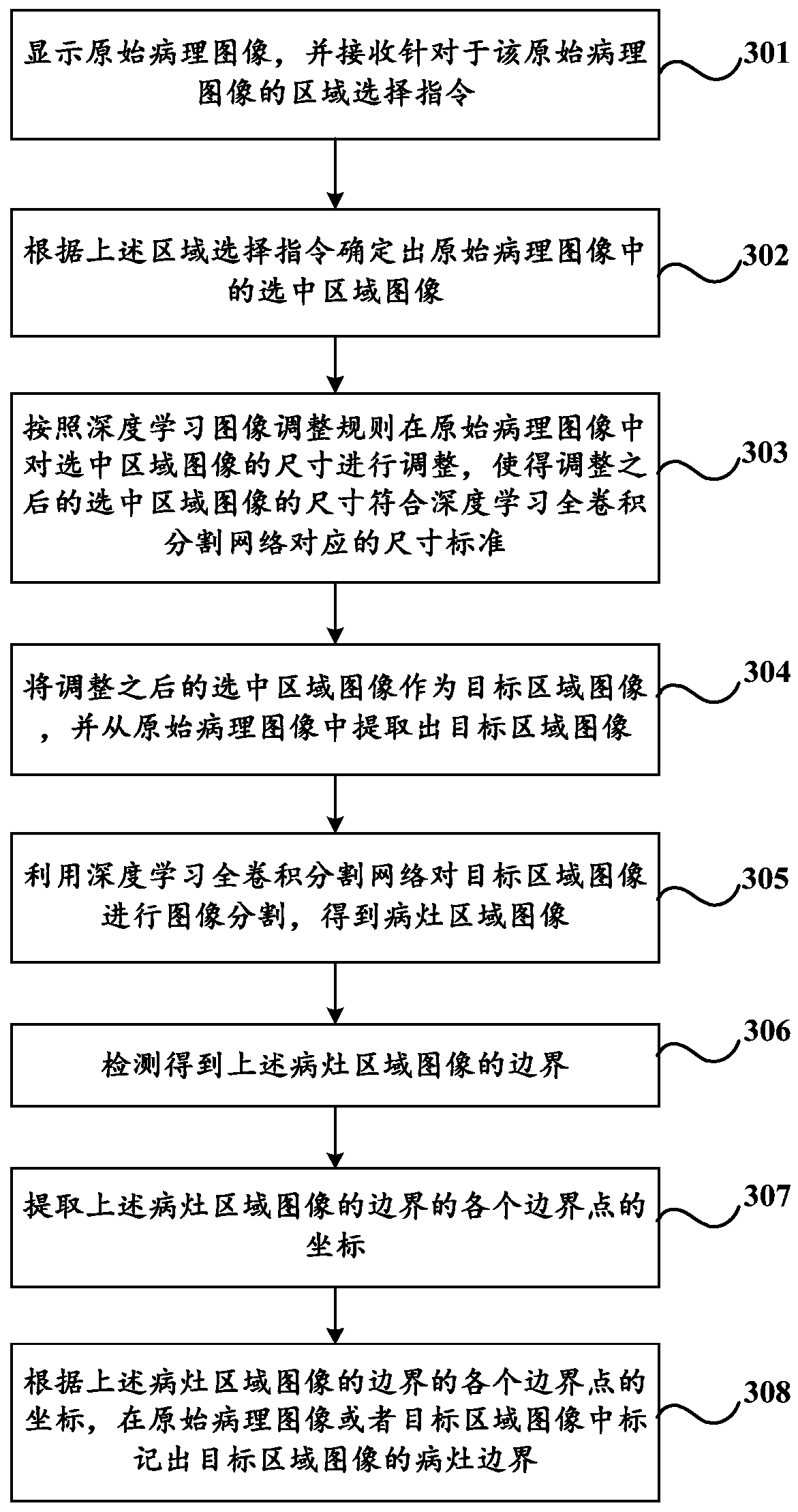
The invention discloses an image processing method and device and a storage medium. The method comprises the steps of extracting a target area image in an original pathological image; performing imagesegmentation on the target area image to obtain a focus area image, the focus area image comprising a focus area of the target area image; and marking a focus boundary of the target area image in thetarget area image or the original pathological image according to the focus area image. According to the image processing method, after the target area image is extracted from the original pathological image, accurate focus boundary detection can be conducted on the target area image, and therefore the image processing method used for achieving area-level accurate focus boundary detection is provided.

Description

technical field [0001] The present application relates to the field of image processing, and in particular to an image processing method, device and storage medium. Background technique [0002] A pathological image is an image that includes a section of a part of human tissue, and is one of the gold standards and important basis for clinical diagnosis of tumor tissues and cells. The traditional pathological image diagnosis method is for medical staff to manually read the slices to interpret the tissue lesions contained in the pathological image. However, this method is not only time-consuming and labor-intensive, but also relies too much on the clinical experience of medical staff, so there are often cases of diagnostic errors. [0003] In this regard, the computer-aided diagnosis method can be used to realize automatic film reading, that is, the use of machines to process pathological images to identify the tissue lesions contained in the pathological images. This method ...

Claims


Application Information

Patent Timeline
Patent Type & Authority Applications(China)
IPC IPC(8): G06T7/12G06T7/11
CPCG06T7/12G06T7/11G06T2207/30004G06T2207/30096
Inventor 黄晓迪李嘉辉
Owner SHANGHAI SENSETIME INTELLIGENT TECH CO LTD
Learn More