
Fundus image blood vessel segmentation method

A fundus image and blood vessel technology, which is applied in image analysis, image enhancement, image data processing, etc., can solve the problems of complex parameter estimation process in the work implementation steps, and the segmentation accuracy of fundus blood vessels needs to be improved, so as to improve the segmentation accuracy and achieve excellent performance , the effect of reducing computational steps

Active Publication Date: 2019-09-03
SHENZHEN GRADUATE SCHOOL TSINGHUA UNIV
View PDF7 Cites 12 Cited by
  • Summary
  • Abstract
  • Description
  • Claims
  • Application Information

AI Technical Summary

Problems solved by technology

However, the implementation steps of the above work are relatively complicated and rely on a complex parameter estimation process. The accuracy of the segmentation of fundus blood vessels, especially the segmentation accuracy of small blood vessels, needs to be improved.

Method used


Image

  • Fundus image blood vessel segmentation method
  • Fundus image blood vessel segmentation method
  • Fundus image blood vessel segmentation method

Examples


Embodiment Construction

[0036]In order to make the technical problems, technical solutions and beneficial effects to be solved by the embodiments of the present invention clearer, the present invention will be further described in detail below in conjunction with the accompanying drawings and embodiments. It should be understood that the specific embodiments described here are only used to explain the present invention, not to limit the present invention.

[0037] It should be noted that when an element is referred to as being “fixed” or “disposed on” another element, it may be directly on the other element or be indirectly on the other element. When an element is referred to as being "connected to" another element, it can be directly connected to the other element or indirectly connected to the other element. In addition, the connection can be used for both fixing and circuit communication.

[0038] It is to be understood that the terms "length", "width", "top", "bottom", "front", "rear", "left", "...


PUM


Abstract

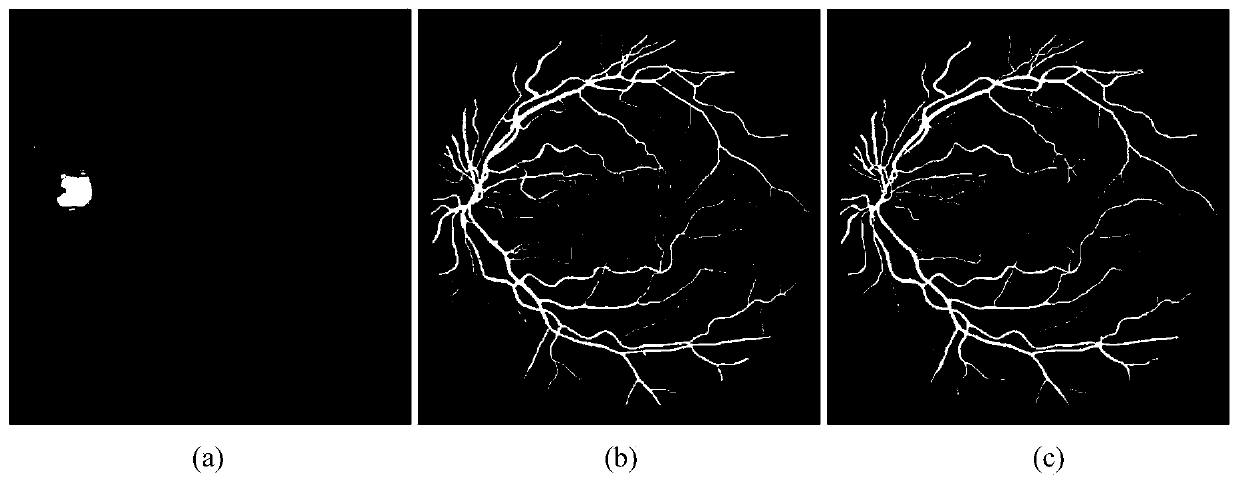
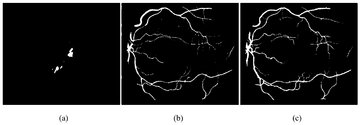
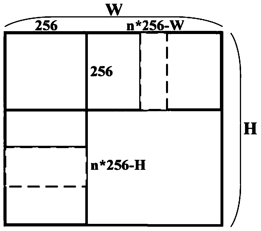
The invention provides a fundus image blood vessel segmentation method which comprises the following steps of S1, carrying out augmented operation on a fundus image data set, namely cutting an original image according to a certain rule to obtain an augmented data set, and dividing the data set into a training set and a test set; S2, carrying out blood vessel segmentation on each fundus image sample in the augmented training data set by using a segmentation network to obtain a black-white probability graph, and carrying out image processing to obtain a probability graph of segmented images in the training data set; S3, using a discrimination network to distinguish whether the probability graph is obtained by a segmentation network or a segmentation image in a data set, and obtaining a confidence graph with a numerical value range of 0-1 to describe the probability that the input image is judged to be true or false; and S4, inputting the color fundus image in the test set into the trained network, and splicing the obtained result images to obtain a complete segmentation image of the fundus blood vessel, wherein the step S2 and the step S3 perform fundus image blood vessel segmentation based on the generative adversarial network, and before the step S4, the segmented network in the step S2 and the judgment network in the step S3 are sequentially subjected to iterative training according to the designed loss function, and the performances of the two networks are respectively improved in a game mode. The method is based on the superiority of the generative adversarial idea in data generation, does not depend on selection of initial features, reduces the complex calculation steps, effectively improves the segmentation precision of fundus image blood vessels, and especially has the excellent performance on tiny blood vessels.

Description

technical field [0001] The invention relates to a non-medical purpose fundus image blood vessel segmentation method. Background technique [0002] As an important part of the eyeball structure, the retina has a very important connection with various systems of the whole body. The retina consists of the optic disc, macula, and retinal blood vessels. As the only deep microvascular system in the human body that can be directly observed by non-invasive means, retinal blood vessels can provide ophthalmologists with important information about changes in their structural state, such as arteriosclerosis, hypertension, diabetes, cardiovascular disease and old age. Macular degeneration, etc. will affect the blood vessels of the fundus, causing changes in the width and curvature of the arteries and veins. This information provides key material for eye-related scientific research. [0003] At present, fundus images are commonly used clinically to observe the status of retinal blood ...

Claims


Application Information

Patent Timeline
IPC IPC(8): G06T7/11G06T5/40G06T3/40G06N3/08G06N3/04
CPCG06T7/11G06T5/40G06N3/08G06T3/4038G06T2207/30041G06N3/045
Inventor 董宇涵任文婷张凯
Owner SHENZHEN GRADUATE SCHOOL TSINGHUA UNIV
Learn More