
Uterus intracavity biopsy extractor

A technology of extractor and uterine cavity implantation, which is applied in the field of gynecological tumor disease treatment, can solve the problems of reducing the work efficiency and work quality of medical staff, high probability of wound infection, and increasing the pain of patients, so as to improve work efficiency and work quality, shrink the The effect of clamping space and reducing pain

Pending Publication Date: 2019-09-17
GUIZHOU PROVINCIAL PEOPLES HOSPITAL
View PDF0 Cites 0 Cited by
  • Summary
  • Abstract
  • Description
  • Claims
  • Application Information

AI Technical Summary

Problems solved by technology

[0003] At present, in the operation of cervical cavity tumors, most of the forceps are used to extend into the uterine cavity to grasp the tumor for biopsy. When the forceps are used to extend into the uterine cavity, the incision of the uterine cavity needs to provide space for the opening and closing of the forceps , leading to large incisions in the uterine cavity, slow recovery of patients after surgery, and a high probability of wound infection, which increases the pain of patients and reduces the work efficiency and quality of medical staff

Method used


Image

  • Uterus intracavity biopsy extractor
  • Uterus intracavity biopsy extractor
  • Uterus intracavity biopsy extractor

Examples


Embodiment approach 1

[0044] The first embodiment of the present invention provides an intrauterine biopsy extractor, which is used to extend into the uterine cavity to clamp and fix the tumor in the uterine cavity of the patient, so as to facilitate the resection of the tumor.

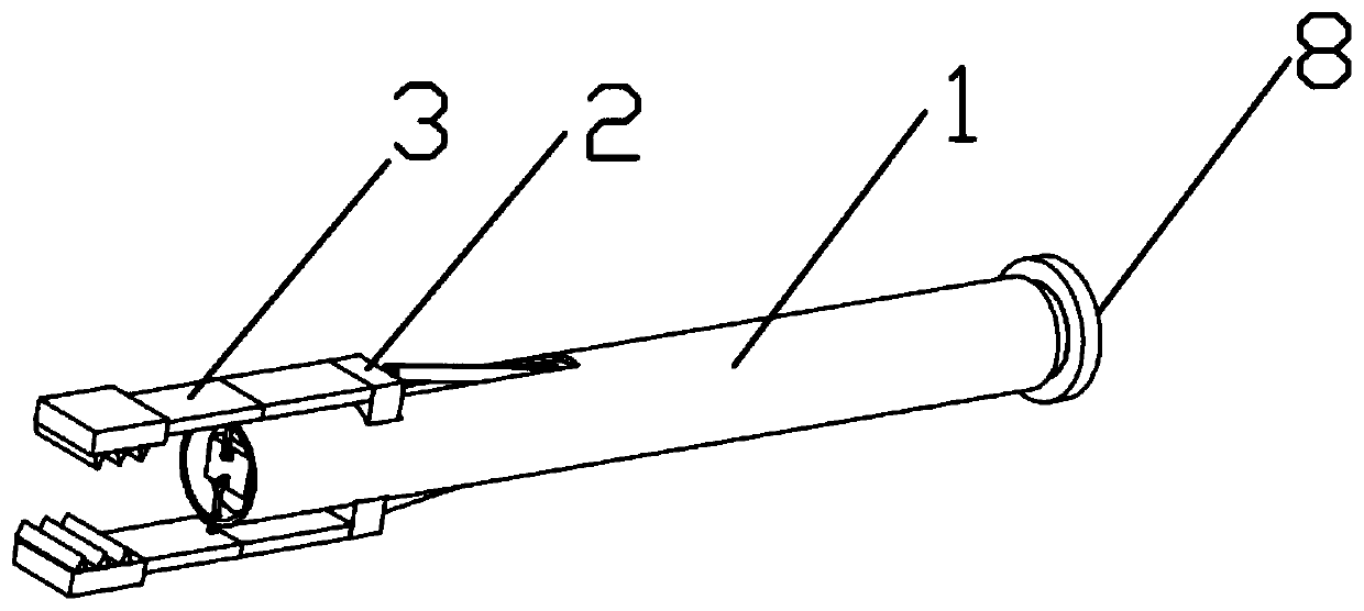
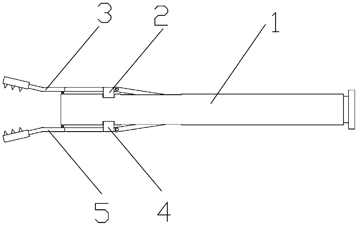
[0045] Such as figure 1 with figure 2 As shown, the intrauterine biopsy extractor includes: a tube body 1, a first lifting member 2, a first clamping member 3, a second lifting member 4, a second clamping member 5, a clamping member 6 and a power assembly 8. The tube body 1 is hollow for extending into the uterine cavity, and the first clamping member 3 and the second clamping member 5 are used to cooperate and clamp the patient's tumor. The power assembly 8 is used to drive the clamping member 6 to push the first lifting member 2 and the second lifting member 4 to expand the clamping space between the first clamping member 3 and the second clamping member 5, so as to clamp larger volume Tumor. Combine Image 6 The intraute...

Embodiment approach 2

[0060] The second embodiment of the present invention relates to an intrauterine biopsy extractor. The second embodiment is basically the same as the first embodiment, except that the intrauterine biopsy extractor in this embodiment can hold a larger volume Tumor.

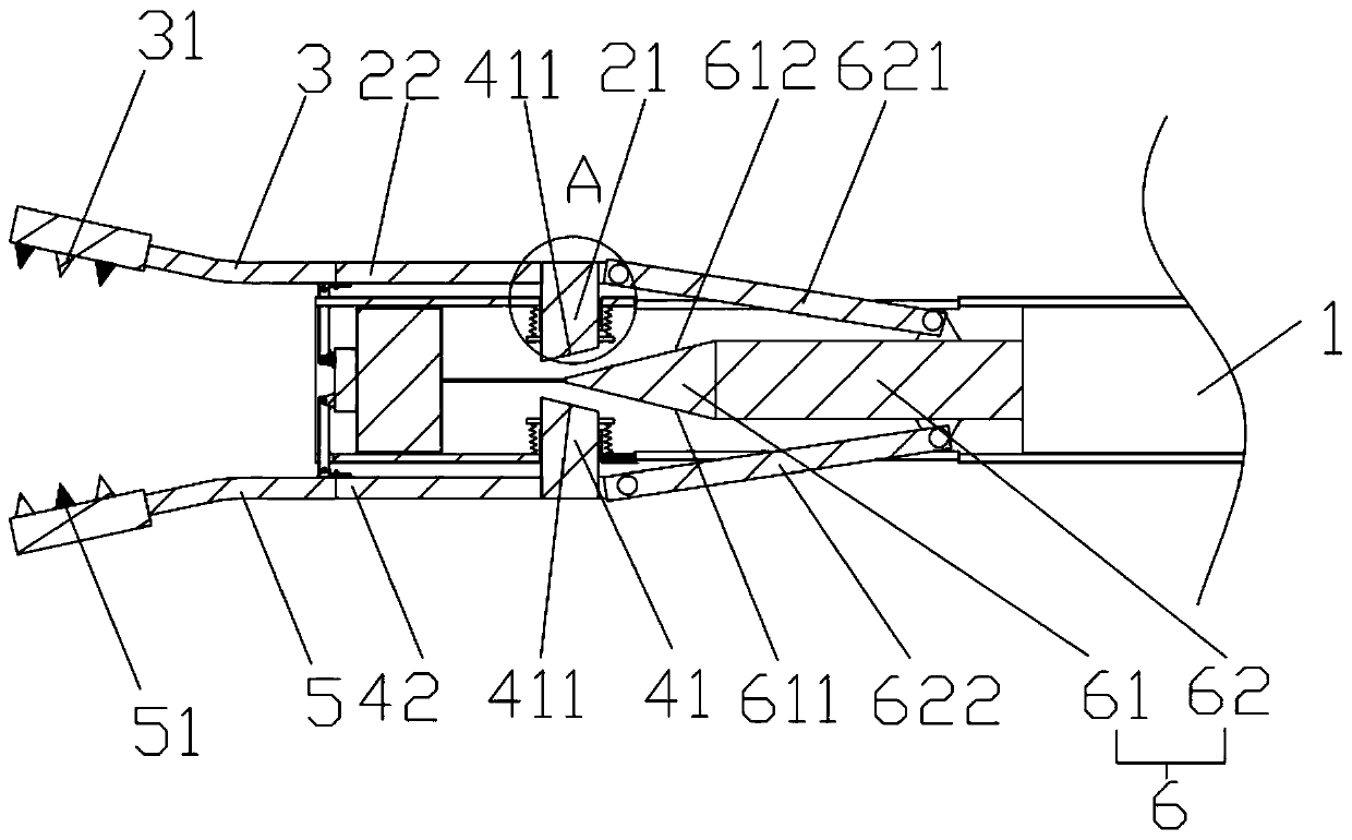
[0061] In this embodiment, as image 3 with Figure 5 As shown, the first lifting rod 21 includes: a first hollow portion 212 penetrating through the side wall of the tube body 1, a first lifting portion 213 arranged in the first hollow portion 212 and slidingly connected to the first hollow portion 212, The sliding direction of the portion 213 is perpendicular to the axial direction of the pipe body 1. The expansion member includes: a pointed head 61 and an expansion member body 62. The expansion member body 62 is connected to the connecting member 82. The expansion member body 62 is provided with a first connecting rod 621. One end of the first connecting rod 621 is connected to the expansion member body. The other...


PUM


Abstract

The invention provides a uterus intracavity biopsy extractor. The uterus intracavity biopsy extractor includes a pipe, a first lifting part, a first clamping part, a second lifting part, a second clamping part, an expansion clamping part and a driving force part; the pipe is used for stretching into the uterine cavity of a patient; the sliding direction of the first lifting part on the pipe is perpendicular to the axle center of the pipe; the first clamping part is hinged to the first lifting part; the sliding direction of the second lifting part on the pipe is perpendicular to the axle center of the pipe; the second clamping part is hinged to the second lifting part and is used for cooperating with the first clamping part to clamp tumors; the expansion clamping part is arranged inside the pipe, is provided with a first slope and a second slope and is used for pushing the first lifting part to slide through the first slope and pushing the second lifting part to slide through the second slope when moving towards the uterine cavity, wherein the first slope is located at the side, facing the first lifting part, of the expansion clamping part, and the second slope is located at the side, facing the second lifting part, of the expansion clamping part; the driving force part is arranged at the side, away from the expansion clamping part, of the pipe and is used for driving the expansion clamping part to move towards or away from the uterine cavity inside the pipe.

Description

Technical field [0001] The invention relates to the field of treatment of gynecological tumor diseases, in particular to an intrauterine biopsy extractor. Background technique [0002] In order to minimize the degree of invasiveness to the patient, in cervical tumor surgery, a biopsy method is generally adopted for the lesion site first, that is, a small piece of diseased tissue is clamped and sent to the pathology department for film production. Determine the benign and malignant, and then decide the follow-up treatment. [0003] At present, in cervical tumor surgery, most of the clamping forceps are used to extend into the uterine cavity to grasp the tumor for biopsy. When the clamping forceps are used to extend into the uterine cavity, the uterine cavity incision needs to provide space for the opening and closing of the grasping forceps. , Resulting in large uterine incisions, slower recovery of patients after surgery, high wound infection probability, increased patient sufferi...

Claims


Application Information

Patent Timeline
Patent Type & Authority Applications(China)
IPC IPC(8): A61B10/02
CPCA61B10/0291
Inventor 张汉群李勇王姿鲁亮杨飞月
Owner GUIZHOU PROVINCIAL PEOPLES HOSPITAL
Learn More