
Liver tumor CT image computer-aided diagnosis method

A computer-aided, CT imaging technology, applied in the field of image processing, can solve the problems of large impact on feature selection and extraction of classification results, and achieve the effects of improving accuracy, simplifying processes, and improving robustness

Active Publication Date: 2019-09-20
SHANGHAI UNIV
View PDF6 Cites 9 Cited by
  • Summary
  • Abstract
  • Description
  • Claims
  • Application Information

AI Technical Summary

Problems solved by technology

More importantly, its classification method is based on image features, resulting in classification results greatly affected by feature selection and extraction

Method used


Image

  • Liver tumor CT image computer-aided diagnosis method
  • Liver tumor CT image computer-aided diagnosis method
  • Liver tumor CT image computer-aided diagnosis method

Examples


Embodiment Construction

[0032] Preferred embodiments of the present invention are described as follows in conjunction with the accompanying drawings:

[0033] Before the model training, it is necessary to prepare the data for training the FCN model and the data of the CNN model respectively. The training data of the FCN includes the original CT image and the segmentation marks of the liver and tumor. The data file format is NIFTI format, and the CT image size is 512× 512 pixels; the training data of CNN includes the original CT image and tumor classification marks, the data file format is DICOM format, and the CT image size is 512×512 pixels.

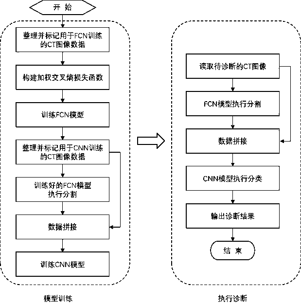
[0034] like figure 1 As shown, a method for computer-aided diagnosis of liver tumor CT images, the operation steps are as follows:

[0035] a) Training the FCN network for liver and tumor segmentation, the specific method is as follows:

[0036] a1) Calculate the vector r of the percentage according to the proportion of the three categories of background, li...


PUM


Abstract

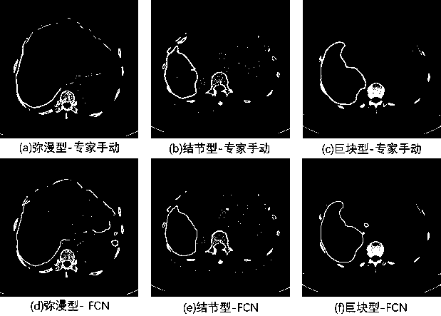
The invention relates to a liver tumor CT image computer-aided diagnosis method. According to the method, the liver and the tumor are segmented through a fully convolutional network (FCN), and the liver tumor is classified through a convolutional neural network (CNN). In training an FCN model, a weighted cross entropy loss function is used for improving tumor segmenting accuracy. In training the CNN and performing classification by means of the CNN, a CT image in a first channel and an FCN segmenting result in a third channel are spliced for obtaining four-channel image data as an input. Finally the trained FCN and the CNN model are combined for constructing a computer-aided diagnosis system. After the to-be-diagnosed CT image is read and input into the system, a probability that the CT image belongs to the healthy liver, the diffuse liver, the tubercle type tumor or the massive cancer is obtained. The integral process of the method does not require the steps of image preprocessing and characteristic extracting. Not only is process simplified, but also the diagnosis accuracy is not affected by image noise, low contrast and characteristic selection and extraction.

Description

technical field [0001] The invention belongs to the technical field of image processing, in particular to a method for computer-aided diagnosis of liver tumor CT images. Background technique [0002] Liver tumors are multiple malignant diseases. In 2018, there were more than 840,000 new patients with liver tumors worldwide, ranking seventh among all types of tumors. Hepatocellular carcinoma is the most common primary liver malignant tumor. It has the characteristics of high malignancy, rapid disease progression, and inconspicuous early symptoms. Once symptoms appear, it is often in the middle and late stages, so the treatment is difficult and the effect is poor. The general survival time after onset is only six months, so it is called "the king of cancer". In the current clinical diagnosis of liver tumors, doctors mostly find lesions through CT images of patients. However, when the CT image quality is not high, the pulse period is single, the doctor is inexperienced or the...

Claims


Application Information

Patent Timeline
IPC IPC(8): G16H50/20G16H30/20
CPCG16H50/20G16H30/20
Inventor 李静吴雨润沈南燕张宇辰孙杰
Owner SHANGHAI UNIV
Learn More