

Pathological auxiliary diagnosis method
A technology for auxiliary diagnosis and pathology, applied in the field of disease diagnosis, can solve the problems of low degree of informatization and the quality of diagnosis needs to be further improved, and achieve the effect of shortening the work process, saving labor costs, and speeding up
- Summary
- Abstract
- Description
- Claims
- Application Information
AI Technical Summary
Problems solved by technology
Method used
Image

Examples
Embodiment Construction
[0041] The following will clearly and completely describe the technical solutions in the embodiments of the present invention with reference to the accompanying drawings in the embodiments of the present invention. Obviously, the described embodiments are only some, not all, embodiments of the present invention. Based on the embodiments of the present invention, all other embodiments obtained by persons of ordinary skill in the art without creative efforts fall within the protection scope of the present invention.
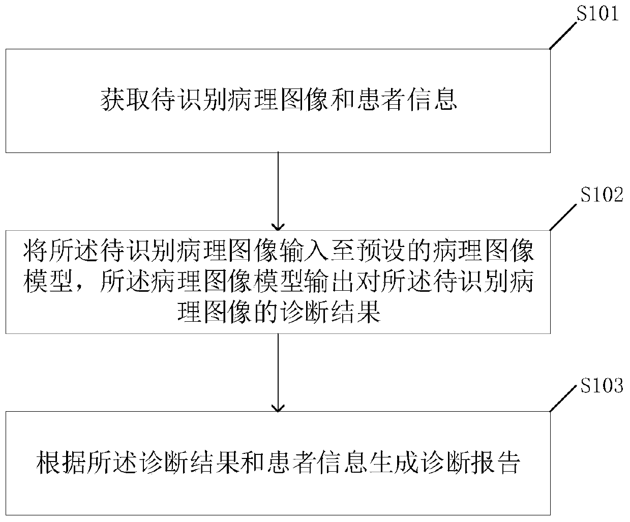
[0042] figure 1 It is a flow chart of the pathological auxiliary diagnosis method provided by the embodiment of the present invention, and the method includes:
[0043] S101. Obtain pathological images to be identified and patient information;
[0044] The pathological image is also called a pathological electronic slice image, which refers to an image scanned by an electron microscope for pathological analysis, such as: a pathological image of the stomach, a path...
PUM
 Abstract
Description
Claims
Application Information