Protein biomarker for assisting in authenticating Rituximab drug-resistant ABC-DLBCL cells and application thereof
What is AI technical title?
AI technical title is built by Patsnap AI team. It summarizes the technical point description of the patent document.
A protein marker and marker technology, applied in animal cells, tumor/cancer cells, vertebrate cells, etc., can solve the problems of poor prognosis and the inability to obtain complete remission with conventional second-line regimens.
Inactive Publication Date: 2019-11-08
CANCER INST & HOSPITAL CHINESE ACADEMY OF MEDICAL SCI
View PDF2 Cites 2 Cited by
Summary
Abstract
Description
Claims
Application Information
AI Technical Summary
This helps you quickly interpret patents by identifying the three key elements:
Problems solved by technology
Method used
Benefits of technology
Problems solved by technology
Although rituximab combined with R-CHOPchemotherapy can significantly improve the outcome of DLBCL patients (the overall cure rate is about 60%), the prognosis of first-line treatment of ABC-DLBCL patients is poor (long-term progression-free survival is less than 50%), and the first-line In patients with drug resistance, more than 70% of conventional second-line regimens cannot achieve complete remission
Method used
Image
Click on the blue label to locate the original text in one second.
Reading with bidirectional positioning of images and text.
Examples
Embodiment 1
[0075] Embodiment 1, the construction of drug-resistant cell line
[0078] 2. After completing step 1, centrifuge at 1000 rpm for 5 minutes, discard the supernatant, add new RPMI-1640 medium containing 1 μg / mL Rituximab and 10% fetal bovine serum, and incubate for 24 hours.
[0079] 3. After completing step 2, centrifuge at 1000rpm for 5min, discard the supernatant, add new RPMI-1640 medium containing 10% fetal bovine serum, and culture the cells to the logarithmic growth phase.
[0080] 4. After completing step 3, centrifuge at 1000 rpm for 5 minutes, discard the supernatant, add new RPMI-1640 medium containing 1 μg / mL Rituximab and 10% fetal bovine serum, and incubate for 24 hours.
[0081] 5. After completing step 3, centrifuge at 1000rpm for 5min, discard the s...
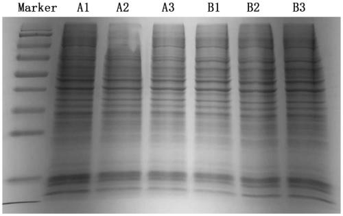
[0109] Take cell samples, add SDT lysate, sonicate (80W, work for 10s, pause for 15s, cycle 10 times), then boil in water bath for 15min, then centrifuge at 14000g for 40min, take the supernatant, which is the protein solution.
[0111] Protein quantification was carried out by BCA method (BCA quantification kit, P0012, Biyuntian). Aliquot the samples and store at -80°C.
[0112] 500 μL protein solution was obtained for each cell sample. Acco...
PUM
Abstract
The present invention discloses a protein biomarker for assisting in authenticating Rituximabdrug-resistant ABC-DLBCL cells and application thereof. The application of a substance for detecting a specific biomarker to preparing a kit for detecting whether to-be-detected ABC-DLBCL cells are Rituximabdrug-resistant ABC-DLBCL cells or not is protected. A biomarker A-1 is any one or a combination of8 proteins. A biomarker A-2 is any one or a combination of 45 proteins. A biomarker B-1 is any one or a combination of 5 proteins. A biomarker B-2 is any one or a combination of 77 proteins. A biomarker C is any one or a combination of 21 proteins. A biomarker D is any one or a combination of 29 proteins. According to the present invention, Rituximabdrug-resistant cell strains are successfully constructed, and multiple protein biomarkers are found. The cell model and research results provide a basis for further studying Rituximab drug-resistance and overcoming drug-resistance.
Description
technical field [0001] The invention relates to a protein marker for assisting identification of Rituximab drug-resistant ABC-DLBCL cells and application thereof. Background technique [0002] Non-Hodgkin lymphoma (Non-Hodgkin lymphoma) is a common malignant tumor of lymphoid tissue, and it is one of the top 10 causes of cancer mortality. In recent years, the incidence of non-Hodgkin lymphoma in my country has also shown an obvious upward trend. Diffuse large B-celllymphoma (DLBCL) is the most common subtype of non-Hodgkin's lymphoma (NHL), accounting for about 32.5% of newly diagnosed NHL cases each year. and other malignant tumors with great heterogeneity. DLBCL can appear in any organ and any part of the body, and the clinical manifestation is an aggressive course. According to different cell origins, DLBCL can be divided into activated B-cell-like DLBCL (ABC-DLBCL), germinal center B-cell-like DLBCL (GCB-DLBCL) and unclassified type. ABC-DLBCL has a poor prognosis and...
Claims
Application Information
Patent Timeline
Application Date:The date an application was filed.
Publication Date:The date a patent or application was officially published.
First Publication Date:The earliest publication date of a patent with the same application number.
Issue Date:Publication date of the patent grant document.
PCT Entry Date:The Entry date of PCT National Phase.
Estimated Expiry Date:The statutory expiry date of a patent right according to the Patent Law, and it is the longest term of protection that the patent right can achieve without the termination of the patent right due to other reasons(Term extension factor has been taken into account ).
Invalid Date:Actual expiry date is based on effective date or publication date of legal transaction data of invalid patent.
Patent Type & AuthorityApplications(China)
IPC IPC(8): G01N33/569G01N33/68C12N5/09
CPCG01N33/56966G01N33/6848C12N5/0693C12N2501/06
Inventor车轶群王迪刘鹏罗扬张岳沈迪曲媛王爽
OwnerCANCER INST & HOSPITAL CHINESE ACADEMY OF MEDICAL SCI

