A Deep Neural Network Algorithm for Automatic Segmentation of Colon Gland Images

A technology of deep neural network and automatic segmentation, which is applied in biological neural network models, image analysis, neural learning methods, etc., can solve the problems of jagged edges of segmented images, cumbersome tasks, and failure to consider the feature information of gland contours, etc., to achieve Efficacy for alleviating imbalance issues, fast auto-segmentation, and improving instance segmentation results

Active Publication Date: 2022-02-01
梅礼晔
View PDF6 Cites 0 Cited by
  • Summary
  • Abstract
  • Description
  • Claims
  • Application Information

AI Technical Summary

Problems solved by technology

Although these methods have achieved good performance on the gland segmentation task in colon tissue, they have not considered the contour feature information of the gland.
The CNN sliding window method is based on image blocks, resulting in jagged edges of the segmented image; the method based on the total variational neural network uses multiple models for prediction, which has the disadvantages of cumbersome tasks and non-sharing of feature information

Method used


Image

  • A Deep Neural Network Algorithm for Automatic Segmentation of Colon Gland Images
  • A Deep Neural Network Algorithm for Automatic Segmentation of Colon Gland Images
  • A Deep Neural Network Algorithm for Automatic Segmentation of Colon Gland Images

Examples


Embodiment 1

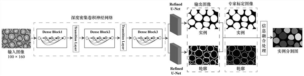
[0022] The present invention will be described in further detail in conjunction with the accompanying drawings and embodiments. In this embodiment, the colon gland image is realized by the automatic segmentation method described in the technical solution for image segmentation, and the specific steps are as follows:

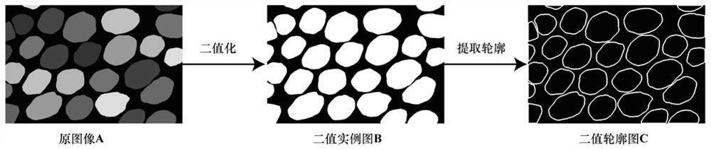
[0023] Step 1: Build the dataset

[0024] Binarize the original Ground Truth image A in the data to obtain a binary instance image B with pixels 0 and 1 as its instance Ground Truth; then use the maximum connected domain method to find the instance contour, and use The morphological expansion extracts the contour and expands it (the structural element dis is 3) to obtain a binary contour map C, which is used as the contour Ground Truth. In order to save memory consumption, in the experiment, the training set data set was scaled to an image of size 100×160, and the training image was standardized by Z-score to ensure the comparability between data. Due to the in...

Embodiment 2

[0069] This embodiment differs slightly from Embodiment 1 only in the evaluation index of the detection model. This embodiment uses specific experimental results and data to illustrate the excellent technical effect of this technical solution.

[0070] Select some experimental results in the MICCAI2015 competition, and the methods of LIST et al., FCN, and CNNTV for comparison. As shown in Table 1, the evaluation indicators comparison of different methods on test set A and test set B. The realization shows that in the test set A, the BF score completely exceeds the current mainstream algorithm, and the Object Dice score and the Object Hausdorff distance score are better than most methods, but on the test set B, all the objective and quantitative evaluation indicators of the algorithm of the present invention completely surpass the current The mainstream method, comprehensive analysis, compared with the current mainstream algorithm, the algorithm of the present invention has cer...


PUM


Abstract

The invention belongs to the technical field of image analysis area segmentation, and discloses a deep neural network algorithm for automatic segmentation of colon gland images. 1. Construct the data set: the colon gland data is used as a training set to obtain the instance map and contour map; 2. Construct the model: the model network includes a dense convolutional neural network and Refined U-Net, and the dense convolutional neural network is used to extract images. Rich primary feature information, the Refined U‑Net network connected with the dense convolutional neural network, learns the feature information of instances and contours; 3. Determine the model loss function: the loss function is the sum of Jaccard and focal loss; 4. Information fusion. This technical solution adopts the characteristics of deep dense neural network feature reuse and high parameter efficiency, and uses the combination of low-level features and high-level features of the Refined U-net network to construct a deep learning network model; uses the focal loss function to solve the problem of data sets Class imbalance problem, effectively make the contour accurate segmentation; finally achieve fast, clear and accurate colon gland image segmentation.

Description

technical field [0001] The invention belongs to the technical field of image analysis area segmentation, and specifically relates to a deep learning-based modeling, a deep dense convolutional neural network and a Refined U-net network. The focus loss function is used as the loss function of the contour task, and the Jaccard distance (Jaccard) is used. As the loss function of the instance segmentation task, Boundary F1 score (Boundary F1, BF), object-level balanced F (Object balanced F, Object F1) score, object-level similarity coefficient (Object Dice), object-level Hausdorff distance (Object Hausdorff ) as a deep neural network algorithm for automatic segmentation of colonic gland images as a quantitative evaluation method. Background technique [0002] The problem of biomedical image segmentation is one of the important research topics for intelligent medical diagnosis tasks, and it is also a difficult point in clinical practice evaluation. In practice, segmentation is us...

Claims


Application Information

Patent Timeline
Patent Type & Authority Patents(China)
IPC IPC(8): G06T7/12G06T5/00G06N3/04G06N3/08
CPCG06T7/12G06T5/005G06N3/08G06T2207/20036G06T2207/30028G06N3/045
Inventor 郭晓鹏梅礼晔孟令玉李华光
Owner 梅礼晔
Why Eureka
Try Eureka