Method for reconstructing cell dynamic characteristic three-dimensional image under biological microscope

A microscopic and dynamic feature technology, applied in the field of medical microscope image processing, can solve the problems of inability to analyze cell structure and morphology, and cannot meet the goals of high-quality diagnostic work, and achieve the effect of solving dynamic problem analysis

Active Publication Date: 2019-11-22
ZHEJIANG SHUREN UNIV
View PDF5 Cites 5 Cited by
  • Summary
  • Abstract
  • Description
  • Claims
  • Application Information

AI Technical Summary

Problems solved by technology

Currently, the functions of the blood cell analyzers used in medical institutions in my country have yet to be improved, and the cells themselves also have diverse morphological characteristics. It may not be possible to conduct in-depth analysis of the specific structure and shape of the cells through instrument detection.
Therefore, it can be concluded that the analysis of patients' blood cell samples by means of instrumental detection alone cannot meet the goal of high-quality diagnosis. The results of manual microscope inspection can further improve the accuracy of diagnosis work, and the reconstructed image of the microscope can play a role at this time

Method used


Image

  • Method for reconstructing cell dynamic characteristic three-dimensional image under biological microscope
  • Method for reconstructing cell dynamic characteristic three-dimensional image under biological microscope
  • Method for reconstructing cell dynamic characteristic three-dimensional image under biological microscope

Examples


Embodiment Construction

[0061] The present invention will be described in further detail below in conjunction with the accompanying drawings and specific embodiments.

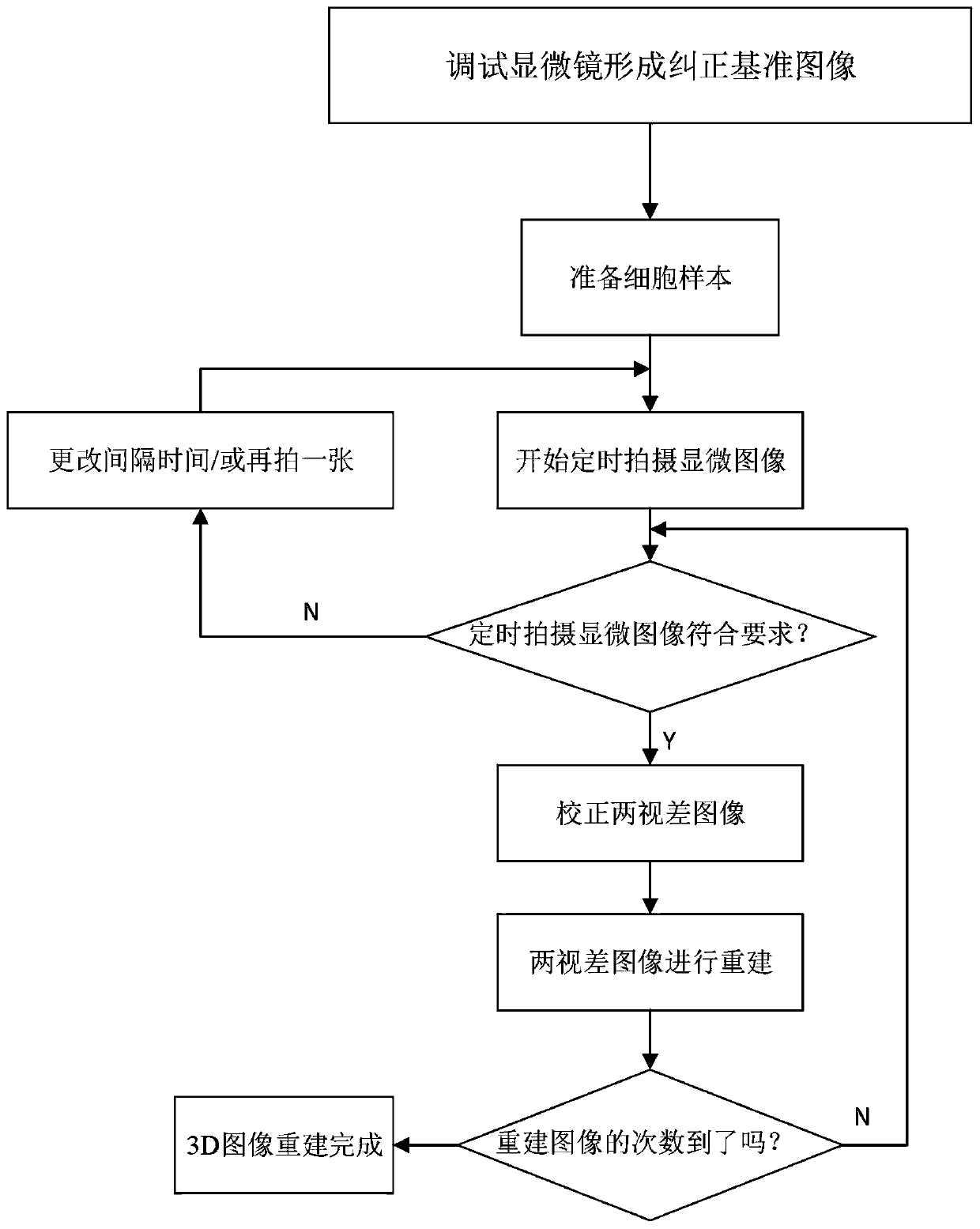
[0062] Such as figure 1 As shown, the present invention provides a method for three-dimensional image reconstruction of dynamic characteristics of cells under a biological microscope, comprising the following steps:

[0063] Step 1: Acquire the reference image of the optical microscope, calculate the anisotropy feature of the brightness gradient for each local area of ​​the acquired microscopic image, scan the local area, obtain the anisotropy distribution map of the area, and then create a An anisotropy map, that is, the reference image of the optical microscope, any optical system itself has errors, that is, system errors. In order to obtain the best optical image of the system, the microscopic image needs to be corrected, so it is necessary to obtain the reference image of the microscopic image, In order to obtain more accurate mi...


PUM


Abstract

The invention provides a method for reconstructing a cell dynamic characteristic three-dimensional image under a biological microscope. The method comprises the following steps: 1, acquiring a reference image of an optical microscope; 2, debugging the optical axis of an optical microscope to a central line, preparing a cell sample, and shooting a cell microscopic image at regular time; and 3, carrying out three-dimensional reconstruction on the two shot qualified two-dimensional cell microscopic images. The implementation method of the step 3 specifically comprises the following steps: (1) dynamic target segmentation and position calculation; (2) constructing a disparity map; and (3) calculating a dynamic cell image by utilizing the three-dimensional characteristics. According to the method, a three-dimensional dynamic object segmentation method is used, the optical axis and correction of deviation are determined, and the 3D characteristics of cells are estimated by combining the disparity map and the 2D characteristics of the initial image, so that the three-dimensional image reconstruction of the dynamic characteristics of the cells is completed. According to the method, the actual value of the distance and the contour of the cell can be quickly determined, the values of all source information are reserved, and the dynamic problem analysis is effectively solved.

Description

technical field [0001] The invention belongs to the technical field of medical microscope image processing, and in particular relates to a method for reconstructing a three-dimensional image of dynamic characteristics of cells under a biological microscope. Background technique [0002] Cells are the basic unit of life, and the study of life needs to start with cells. Ordinary biological microscopy laboratories in colleges and universities can do cell research, but the workload of manual observation of cell number and shape is too large. Since the emergence of modern computers, computer image processing technology can be used to modify existing optical microscopy laboratories to realize The research of cells is automated, which also saves a lot of experimental funds and makes full use of existing laboratory resources. [0003] Blood cell morphology examination is an effective means of diagnosing diseases and one of the important contents of routine blood examination. In re...

Claims


Application Information

Patent Timeline
Patent Type & Authority Applications(China)
IPC IPC(8): G06T17/00G06T7/00G06T7/136
CPCG06T17/00G06T7/0012G06T7/136G06T2207/10061G06T2207/30024
Inventor 叶时平白直灿陈华锋陈超祥许娅芬奥尔佳·涅茨韦德谢尔盖·阿布拉梅科亚历山大·涅茨韦德
Owner ZHEJIANG SHUREN UNIV
Why Eureka
Try Eureka