
MRI glioma segmentation method and system based on sparse Bayesian model and multi-map fusion

A sparse Bayesian and glioma technology, applied in the field of medical image processing, can solve problems such as low contrast, long processing time, and high computational consumption, and achieve precise segmentation and clear boundaries

Active Publication Date: 2019-11-26
SHANDONG NORMAL UNIV
View PDF9 Cites 0 Cited by
  • Summary
  • Abstract
  • Description
  • Claims
  • Application Information

AI Technical Summary

Problems solved by technology

Although there are many ways mentioned above in the segmentation of brain tumors, however, the inventor found in the course of the research that none of the ways can well solve the problem of low contrast between the tumor area and the adjacent region of interest
In addition, excessive processing time and high computational consumption are not acceptable in clinical practice

Method used


Image

  • MRI glioma segmentation method and system based on sparse Bayesian model and multi-map fusion
  • MRI glioma segmentation method and system based on sparse Bayesian model and multi-map fusion
  • MRI glioma segmentation method and system based on sparse Bayesian model and multi-map fusion

Examples


Embodiment 1

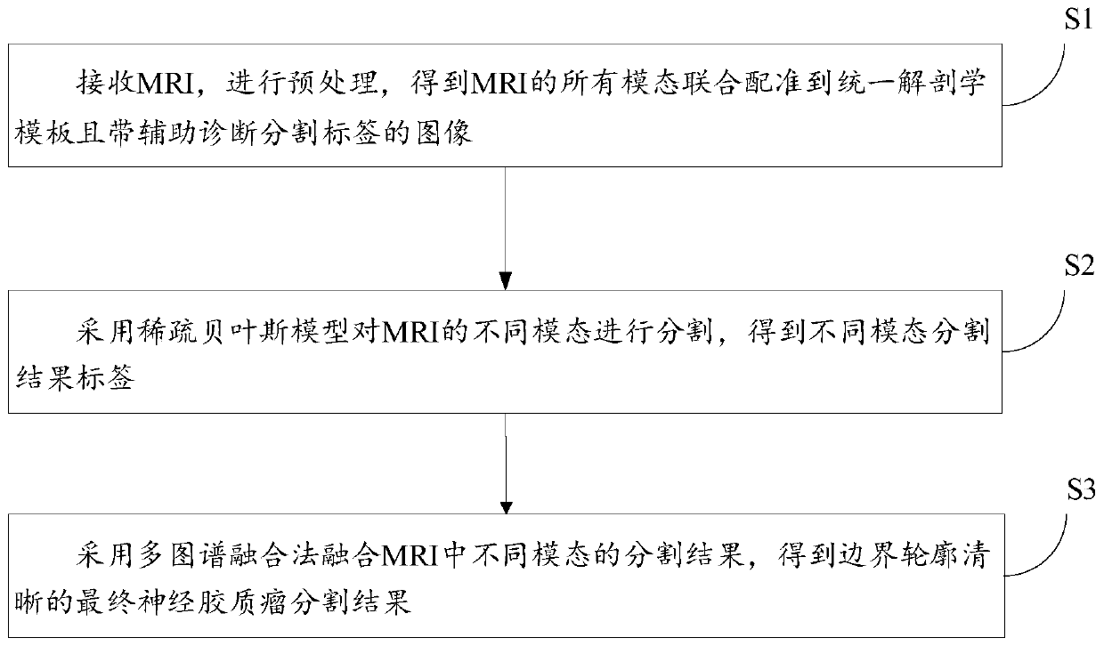
[0045] Such as figure 1 As shown, an MRI glioma segmentation method based on sparse Bayesian model and multi-atlas fusion, the method includes:

[0046] An MRI glioma segmentation method based on sparse Bayesian model and multi-atlas fusion, the method includes:

[0047] Step S1: Receive the MRI, perform preprocessing, and obtain an image in which all modalities of the MRI are co-registered to a unified anatomical template and have segmentation labels for auxiliary diagnosis;

[0048] The sparse Bayesian model is used to segment the different modalities of MRI, and the segmentation result labels of different modalities are obtained;

[0049] The multi-atlas fusion method was used to fuse the segmentation results of different modalities in MRI to obtain the final glioma segmentation results with clear boundaries.

[0050] In step S1 of one or more embodiments of the present disclosure, the specific steps of the preprocessing include:

[0051] The GLISTRboost algorithm is use...

Embodiment 2

[0083] According to an aspect of one or more embodiments of the present disclosure, there is provided a computer-readable storage medium. A computer-readable storage medium, wherein a plurality of instructions are stored, and the instructions are suitable for being loaded by a processor of a terminal device and executing the MRI glioma based on a sparse Bayesian model and multi-atlas fusion split method.

Embodiment 3

[0085] According to an aspect of one or more embodiments of the present disclosure, a terminal device is provided.

[0086] A terminal device, which includes a processor and a computer-readable storage medium, the processor is used to implement instructions; the computer-readable storage medium is used to store multiple instructions, and the instructions are suitable for being loaded by the processor and executing the described one An MRI Glioma Segmentation Method Based on Sparse Bayesian Model and Multi-Atlas Fusion.

[0087] These computer-executable instructions, when executed in a device, cause the device to perform the methods or processes described in accordance with various embodiments in the present disclosure.

[0088]In this embodiment, a computer program product may include a computer-readable storage medium carrying computer-readable program instructions for performing various aspects of the present disclosure. A computer readable storage medium may be a tangible...


PUM


Abstract

The invention discloses an MRI glioma segmentation method and system based on a sparse Bayesian model and multi-map fusion, and the method comprises the steps: receiving MRI, carrying out the preprocessing, and obtaining an image which is jointly registered to a unified anatomy template through all modes of the MRI and is provided with an auxiliary diagnosis segmentation label; segmenting different modes of the MRI by adopting a sparse Bayesian model to obtain different mode segmentation result tags; and fusing segmentation results of different modes in MRI by adopting a multi-map fusion method to obtain a final glioma segmentation result with a clear boundary contour.

Description

technical field [0001] The disclosure belongs to the technical field of medical image processing, and relates to an MRI glioma segmentation method and system based on a sparse Bayesian model and multi-atlas fusion. Background technique [0002] The statements in this section merely provide background information related to the present disclosure and do not necessarily constitute prior art. [0003] In recent years, with the development of medical technology, medical image processing has become more and more important, and the segmentation of brain tumors based on MRI images has become a hot topic. The segmentation of MRI images with tumors and the extraction of tumor information provide important reference value for disease diagnosis, surgical plan formulation, and disease tracking. The task of glioma segmentation is to distinguish normal tissue from diseased tissue in MRI images with tumors, and to make the boundaries of different sub-regions of the tumor clear. However, ...

Claims


Application Information

Patent Timeline
Patent Type & Authority Applications(China)
IPC IPC(8): G06T7/11G06K9/62G06T7/174
CPCG06T7/11G06T7/174G06T2207/10088G06T2207/30096G06F18/24155G06F18/25
Inventor 王晶晶赵兴昊许化强张立人
Owner SHANDONG NORMAL UNIV
Learn More