Living pig carcass tissue content determination method based on CT image processing

A technology of CT image and measurement method, which is applied in the field of image processing, and can solve problems such as unsuitable determination of artiodactyl tissue content

Active Publication Date: 2019-12-24
史记生物技术(南京)有限公司 +1
View PDF6 Cites 6 Cited by
  • Summary
  • Abstract
  • Description
  • Claims
  • Application Information

AI Technical Summary

Problems solved by technology

This application can provide technical support for the quantitative analysis of blood vessels in the lower limbs of living animals, but the quantitative

Method used


Image

  • Living pig carcass tissue content determination method based on CT image processing
  • Living pig carcass tissue content determination method based on CT image processing
  • Living pig carcass tissue content determination method based on CT image processing

Examples


Embodiment 1

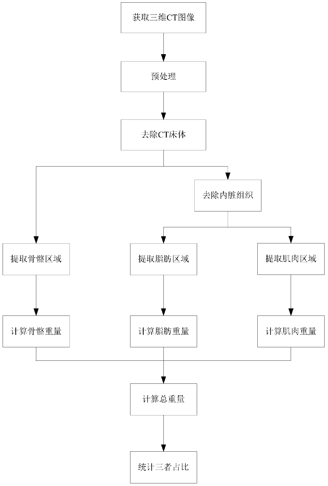
[0081] Such as figure 1 As shown, the method for measuring the content of live pig carcass tissue based on CT image processing in this embodiment includes the following steps:

[0082] (1) Obtain a CT scan image, and perform Gaussian convolution smoothing and denoising preprocessing on the CT scan image; which specifically includes the following steps:

[0083] (11) Extract the CT image information, obtain the two-dimensional slices of the CT images of breeding pigs, and superimpose the two-dimensional slices in order to generate a three-dimensional matrix f(x, y, z). The three-dimensional display of the CT image is as follows figure 2 As shown, the cross-sectional, sagittal, and coronal views of the CT image are as follows image 3 Shown

[0084] (12) Preprocess the acquired CT image, use Gaussian filtering to denoise the image, and Gaussian convolution denoising is as follows:

[0085] g(x,y,z)=f*G σ

[0086] Where * Indicates convolution, σ is the standard deviation, taking 0.8, (...


PUM


Abstract

The invention discloses a living pig carcass tissue content determination method based on CT image processing. The invention belongs to the technical field of image processing. The method comprises six steps: 1, obtaining a CT image, and carrying out the denoising of the CT image; 2, segmenting and removing a bed board part in the CT image; 3, extracting a main skeleton part area of the CT image,and calculating the mass and proportion of the main skeleton part area; 4, extracting a main fat part region of the CT image, and calculating the mass and proportion of the main fat part region; 5, extracting and removing the viscera part of the CT image main body; and 6, extracting a main muscle part region of the CT image, and calculating the mass and proportion of the main muscle part region. Based on the characteristics on the CT image, targeted image region segmentation and correlation calculation are adopted, all visceral tissues of the pig carcass can be accurately and rapidly removed at a time, and the weight and proportion of bones, muscles and adipose tissues of the pig carcass are accurately segmented and measured.

Description

Technical field [0001] The invention belongs to the technical field of image processing, and specifically relates to a CT image segmentation and determination algorithm, in particular to a method for determining the content of live pig carcass tissue based on CT image processing. Background technique [0002] Computed Tomography (CT) is a product of the combination of computer and X-ray examination technology. Its principle is to reflect the density of substances according to the degree of absorption of X-ray by various tissues of the body. It is a non-hazardous organ imaging method in vitro one. CT was first used in clinical medicine and achieved certain achievements. With the development of computer technology and material level, the application field of CT has been greatly expanded. In agricultural production, the combined application of CT and computer technology to determine the content of each tissue in vivo has become an important research direction in the current field....

Claims


Application Information

Patent Timeline
IPC IPC(8): G06T7/11G06T5/00G06T7/136G06T7/155
CPCG06T7/11G06T7/136G06T7/155G06T5/002G06T2207/10081Y02P60/87
Inventor 傅衍邰伟鹏潘祥吕成军汪杰李浩
Owner 史记生物技术(南京)有限公司
Why Eureka
Try Eureka