Detection method of transformed DNA recovery rate and primers

A recovery rate and primer pair technology, applied in biochemical equipment and methods, microbial determination/inspection, etc., can solve the problems of lack of mutual confirmation of different detections

Inactive Publication Date: 2020-01-10
SINGLERA HEALTH TECH SHANGHAI LTD
View PDF4 Cites 0 Cited by
  • Summary
  • Abstract
  • Description
  • Claims
  • Application Information

AI Technical Summary

Problems solved by technology

There is currently no easy method to systematically detect the recovery rate of such DNA, and there is a lack of cross-validation between different assays

Method used


Image

  • Detection method of transformed DNA recovery rate and primers
  • Detection method of transformed DNA recovery rate and primers
  • Detection method of transformed DNA recovery rate and primers

Examples


Embodiment 1

[0060] Transformation of embodiment 1-DNA

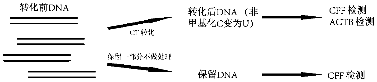
[0061] In this example, the Z kit and the T kit were used to transform DNA. The fragmented reference genome NA12878 DNA was used as the detection DNA, and each kit was repeated 4 times for detection.

[0062] 1) Configure CT conversion reagents according to the instructions of each kit.

[0063]2) Adjust the concentration of fragmented NA12878 to 1 ng / μL, take 20 μL and add 130 μL CT conversion reagent, and repeat the detection 4 times for each kit. Keep about 20 μL of untransformed DNA as a control.

[0064] 3) Run the CT conversion temperature control program according to the instructions of each kit. The Z kit is 98°C for 8 minutes, 54°C for 1 hour, and stored at 4°C. The T kit is 98°C for 10 minutes, 64°C for 2 hours and 30 minutes, and stored at 4°C.

[0065] 4) Purify the post-conversion DNA adsorption column according to the kit instructions, including DNA binding, desulfonation reaction, washing and elution. Finally, 20...

Embodiment 2-C

[0066] Example 2-CFF qPCR detection

[0067] In this example, CFF primers were used to perform qPCR on the transformed DNA to obtain the Ct value.

[0068] 1) Dilute the CFF forward and reverse primers to 4 μM, respectively, take the same volume and mix to form a primer mixture, and configure the qPCR reaction mixture as follows: Kapa SYBR Fast qPCR MIX 5 μL, CFF primer mixture 1 μL, and control or transformed DNA 4 μL.

[0069] CFF forward primer: TAAGAGTAATAATGGATGGATGATG

[0070] CFF reverse primer: CCTCCCATCTCCCTTCC

[0071] 2) Vortex and briefly centrifuge.

[0072] 3) Put the PCR tube into the fluorescent quantitative PCR instrument (BioRad CFX connect Real-Time PCR Detection System), and run the following program:

[0073] Pre-denaturation: 95°C for 5 minutes;

[0074] 40 cycles of signal collection: 95°C for 30 seconds; 54°C for 60 seconds (fluorescence signal collection).

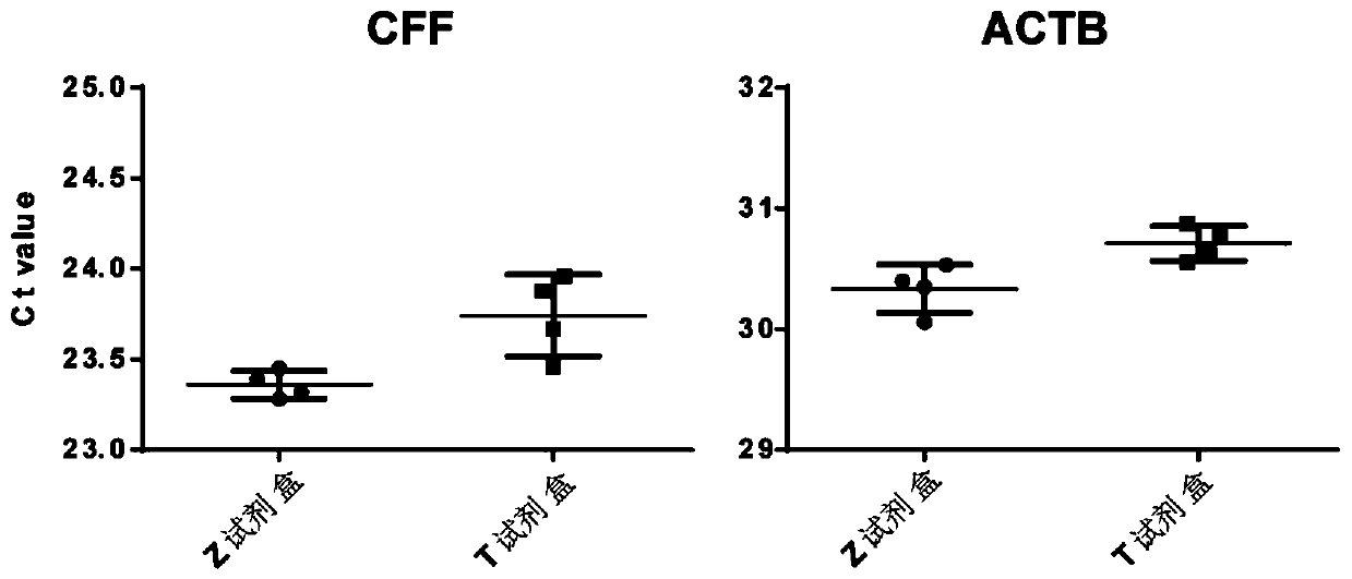
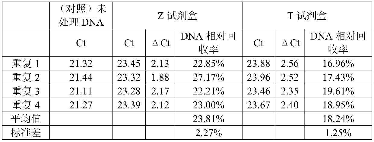
[0075] The results are shown in Table 1 and figure 2 shown. Statistical CFF primer dete...

Embodiment 3-A

[0080] Example 3-ACTB qPCR detection

[0081] In this example, qPCR was performed on the transformed DNA using ACTB primers to obtain the Ct value.

[0082] 1) Dilute the ACTB forward and reverse primers to 10 μM respectively, take the same volume and mix them to form a primer mixture, and dilute the ACTB probe to 4 μM.

[0083] 2) Configure the qPCR reaction as follows: NEB Luna PCR master mix 10 μL, ACTB primer mix 2 μL, ACTB probe 1 μL and transformed DNA 7 μL.

[0084] ACTB forward primer: GTGATGGAGGAGGTTTAGTAAGTT

[0085] ACTB reverse primer: CCAATAAAACCTACTCCCTCCCTAA

[0086] ACTB probe:

[0087] / 5TAMRA / ACCACCACCCAACACACAATAACAAACACA / 3IABkRQ /

[0088] 3) Put the PCR tube into the fluorescent quantitative PCR instrument (ABI 7500 Real-Time PCR System), and run the following program:

[0089] Pre-denaturation: 95°C for 5 minutes;

[0090] 40 cycles of signal collection: 95°C for 15 seconds; 58°C for 60 seconds (fluorescence signal collection).

[0091] The results ...


PUM


Abstract

The present invention provides a detection method of a transformed DNA recovery rate. The detection method comprises the following steps: 1) qPCR is conducted on a DNA sample with primers to obtain atransformed Ct value, wherein the DNA sample is transformed to convert an unmodified cytosine base to a uracil base, and the primers recognize a cytosine base-free DNA fragment in the DNA sample; and2) the Ct value and the primers are used to conduct qPCR of an untransformed DNA sample and the obtained Ct value is used to calculate a DNA recovery rate. The method further comprises conducting qPCRon the transformed DNA sample with the DNA primers recognizing the transformed DNA to obtain a Ct value and then evaluating the DNA recovery rate in the step 2).

Description

technical field [0001] The present invention relates to the field of detection of the recovery of DNA undergoing cytosine conversion. Background technique [0002] Cytosine conversion, such as bisulfite conversion, is the use of bisulfite compounds to treat DNA to convert its unmodified (such as methylated) cytosine base (C) into uracil (U), and then in An in vitro chemical reaction in which methylated cytosines are converted to thymine (T) during subsequent PCR amplification, while the methylated cytosines remain unchanged. After being chemically treated with a bisulfite compound, by qPCR or sequencing detection, it is possible to know which positions of cytosine in the genome are modified by methylation. Therefore, bisulfite conversion is currently the most commonly used DNA treatment method for methylation detection. [0003] In addition to completing the above chemical changes, bisulfite treatment also brings a lot of damage to DNA, resulting in DNA fragmentation and d...

Claims


Application Information

Patent Timeline
Patent Type & Authority Applications(China)
IPC IPC(8): C12Q1/6851
CPCC12Q1/6851C12Q2531/113C12Q2523/125
Inventor 刘蕊王辉
Owner SINGLERA HEALTH TECH SHANGHAI LTD
Why Eureka
Try Eureka