
Tissue mimicking body model for detecting imaging distinguishability of ultrasound echotomography scanning equipment

A technology of tomography and resolution, applied in the field of imitation tissue phantoms, to achieve the effect of original maintainability, increase the validity period of use, and avoid artifacts

Pending Publication Date: 2020-02-07
INST OF ACOUSTICS CHINESE ACAD OF SCI
View PDF0 Cites 2 Cited by
  • Summary
  • Abstract
  • Description
  • Claims
  • Application Information

AI Technical Summary

Problems solved by technology

At present, for USCT, a new ultrasonic diagnostic imaging device, the current tissue-mimicking phantom is no longer suitable for testing its performance

Method used


Image

  • Tissue mimicking body model for detecting imaging distinguishability of ultrasound echotomography scanning equipment
  • Tissue mimicking body model for detecting imaging distinguishability of ultrasound echotomography scanning equipment
  • Tissue mimicking body model for detecting imaging distinguishability of ultrasound echotomography scanning equipment

Examples


Embodiment Construction

[0052] The technical solution of the present invention will be described in detail below in conjunction with the accompanying drawings.

[0053] According to the quality system requirements of medical device manufacturing enterprises and professional quality inspection institutions, all measuring instruments used for quality inspection must be regularly verified or calibrated. Ultrasonic tissue imitation phantoms are "tissue substitutes". Because they directly affect the quality judgment of ultrasonic diagnostic equipment, the rules of regular inspection and comparison have been formed since the end of the last century, which is recognized and followed by relevant circles. The product corresponding to the present invention is used for quality detection and evaluation of ultrasonic tomography equipment, and for detection of resolution of ultrasonic imaging equipment and lesion imaging performance.

[0054] The imitation tissue phantom of the present invention is specially used ...


PUM

PropertyMeasurementUnit
Diameteraaaaaaaaaa

Abstract

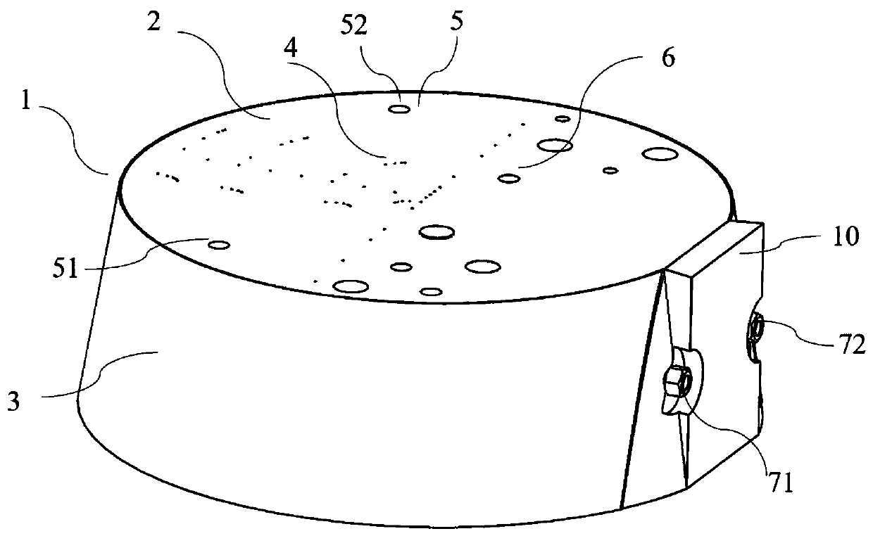
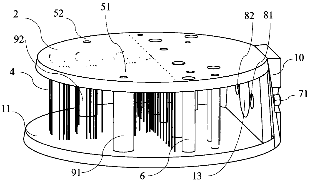
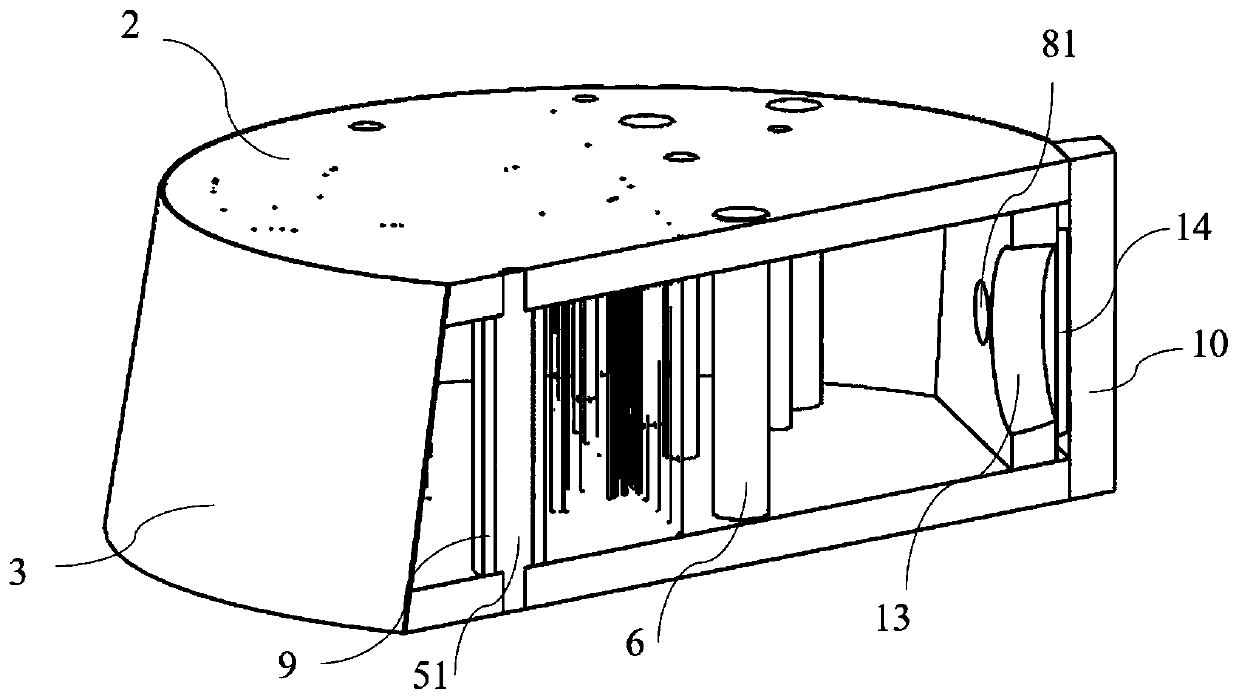
The invention discloses a tissue mimicking body model for detecting imaging distinguishability of ultrasound echotomography scanning equipment. The tissue mimicking body model is in the shape of a truncated cone, and comprises a body model housing, an acoustic window (3), a supporting and protecting plate (6), a plurality of target lines (4), a plurality of simulated niduses (6) and background tissue-mimicking materials (12), wherein the body model housing is manufactured by fixed connecting of an upper panel (2), a lower panel (11) and a supporting plate (14); a sealing space is formed by thebody model housing and the acoustic window (3) which is adhered and fixed to the side surface of the truncated cone; the background tissue-mimicking materials (12) are stuffed in the sealing space; the target lines (4) and the simulated niduses (6) are embedded in the background tissue-mimicking materials (12); each target line and each simulated niduse vertically penetrate from the upper panel (2) to the lower panel (11); an inlet for stuffing the background tissue-mimicking materials (12) is formed in the supporting plate (14); a blocking rubber is adhered to the inlet; and the supporting and protecting plate (6) is fixed outside the supporting plate (14).

Description

technical field [0001] The invention belongs to the field of medical device quality detection, and in particular relates to a tissue imitation phantom used for detection of imaging resolution of ultrasonic tomography equipment. Background technique [0002] B-ultrasound, X-CT, magnetic resonance imaging, and nuclear medicine imaging are the four most practical imaging diagnostic technologies in the contemporary era, and in terms of popularity, B-ultrasound ranks first among the four. Especially under the specific conditions of our country, B-ultrasound technology is not only used in the routine diagnosis of various diseases, but also widely used in the fields of family planning, eugenics and reproductive health. Their performance and quality are not only related to the economic benefits of manufacturers and hospitals, but also affect the health and well-being of the entire Chinese nation, including future generations. It is precisely in view of this situation that the natio...

Claims


Application Information

Patent Timeline
IPC IPC(8): A61B8/00A61B8/08A61B8/13
CPCA61B8/0825A61B8/13A61B8/483A61B8/587
Inventor 张迪牛凤岐朱承纲程洋
Owner INST OF ACOUSTICS CHINESE ACAD OF SCI
Learn More