Method for predicting morphological changes of liver tumor after ablation based on deep learning

A liver tumor and deep learning technology, applied in the field of minimally invasive ablation, can solve problems such as inaccurate evaluation results

Active Publication Date: 2020-02-11
GENERAL HOSPITAL OF PLA
View PDF3 Cites 10 Cited by
  • Summary
  • Abstract
  • Description
  • Claims
  • Application Information

AI Technical Summary

Problems solved by technology

However, no matter which method, in the registration process, only the characteristics of the image itself are considered, and changes such as local tissue shrinkage caused by high temperature during clinical treatment are not taken into account, resulting in inaccurate evaluation results

Method used


Image

  • Method for predicting morphological changes of liver tumor after ablation based on deep learning
  • Method for predicting morphological changes of liver tumor after ablation based on deep learning
  • Method for predicting morphological changes of liver tumor after ablation based on deep learning

Examples


Embodiment Construction

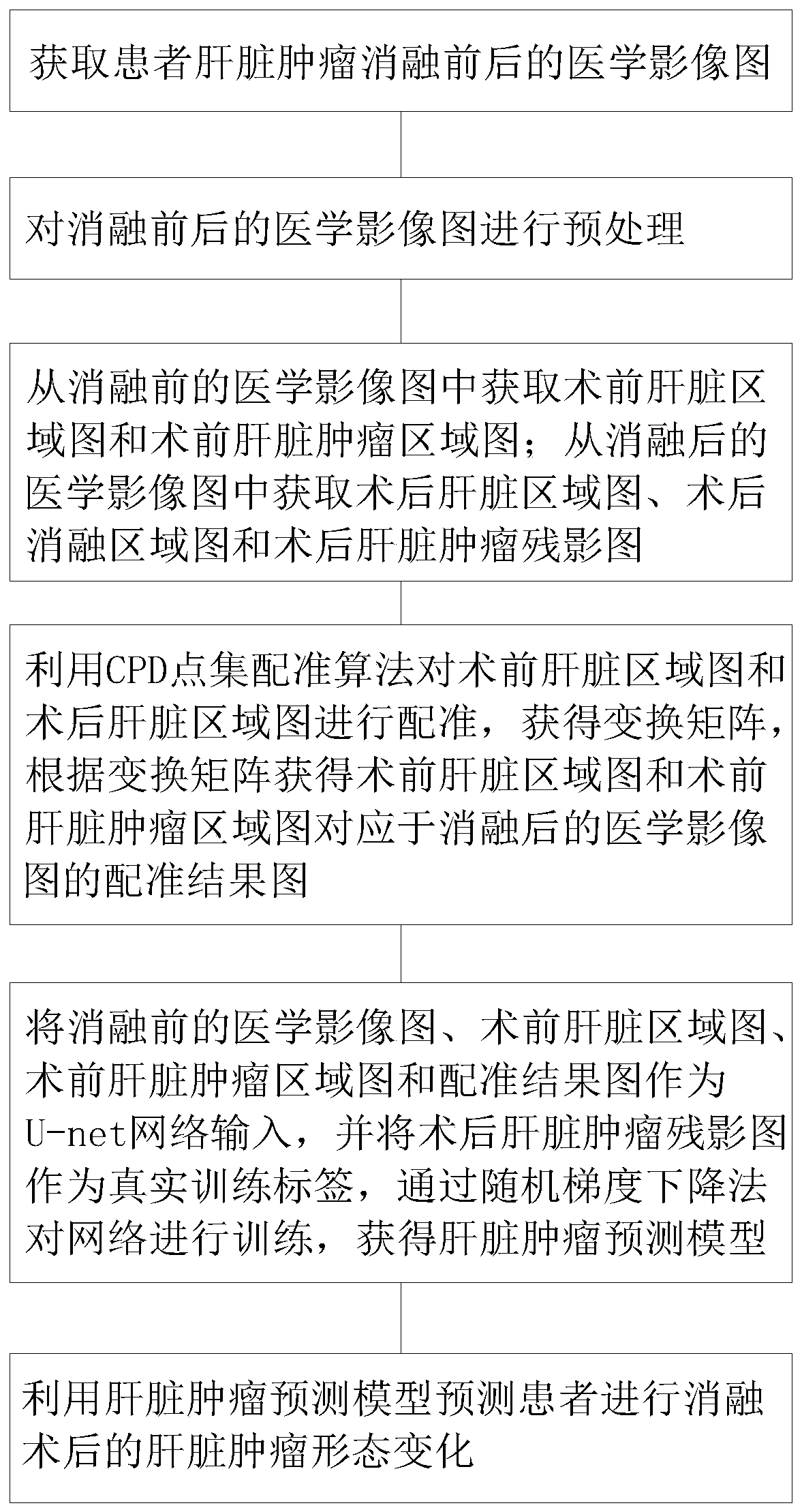
[0042] Such as figure 1 As shown, this embodiment provides a method for predicting the morphological changes of liver tumors after ablation based on deep learning, including the following steps:

[0043] Step 1: Acquire CT / MRI scanning sequence images of the patient's liver tumor before and after ablation.

[0044] Step 2, preprocessing the medical images before and after ablation, specifically: grouping and dividing the data set according to the factors affecting the liver, then reading the CT / MRI scan sequence images before and after ablation, and performing Gaussian analysis on the CT / MRI scan sequence images Denoising, grayscale histogram equalization, image contrast enhancement, rotation, flipping, and data normalization to increase sample diversity while speeding up network convergence. Wherein, the liver influencing factors include factors such as liver status, tumor type and pathological type.

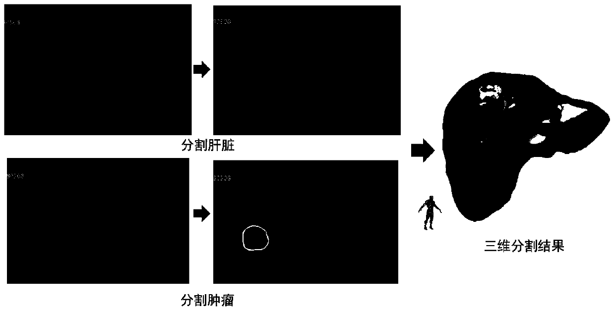
[0045] Step 3: Obtain the preoperative liver region map and preoperative...


PUM


Abstract

The invention discloses a method for predicting the morphological change of a liver tumor after ablation based on deep learning. The method comprises the following steps: acquiring a medical image mapof a patient before and after liver tumor ablation; preprocessing the medical images before and after ablation; obtaining a preoperative liver region map and a preoperative liver tumor region map; acquiring a postoperative liver region map, a postoperative ablation region map and a postoperative liver tumor ghost image; obtaining a transformation matrix by using a CPD point set registration algorithm, and obtaining a registration result graph according to the transformation matrix; training the network through a stochastic gradient descent method to obtain a liver tumor prediction model; andpredicting the morphological change of the liver tumor of the patient after ablation by using the liver tumor prediction model. According to the method, the morphological change of the liver tumor after ablation of the patient can be predicted according to the CT / MRI image of the patient, a basis is provided for quantitatively evaluating whether the ablation area completely covers the tumor, a doctor can accurately evaluate the postoperative curative effect, and a foundation is laid for a subsequent treatment scheme of the patient.

Description

technical field [0001] The invention belongs to the field of minimally invasive ablation, and in particular relates to a method for predicting morphological changes of liver tumors after ablation based on deep learning. Background technique [0002] In recent years, image-guided percutaneous thermal ablation has become one of the most promising minimally invasive treatments for solid tumors such as liver, kidney, and breast. Among them, microwave ablation is guided by ultrasound, CT and other images. The ablation needle is inserted into the tumor, and the release of electromagnetic waves causes local area polar molecules to vibrate and rub to generate high temperature, and finally achieves the purpose of inactivating the tumor. Compared with traditional surgery, it has the advantages of small trauma, good curative effect, fast recovery, repeatability, low cost, and can improve the immune function of the body. It can achieve good curative effect of completely inactivating tum...

Claims


Application Information

Patent Timeline
Patent Type & Authority Applications(China)
IPC IPC(8): G06T7/187G06T7/13G06T7/33G06N3/04
CPCG06T7/187G06T7/13G06T7/33G06T2207/10081G06T2207/10088G06T2207/30056G06T2207/30101G06N3/045G06T7/0012G06T7/344G06T7/11G06T2207/30096G06T2207/20081G06T2207/20076G06T2207/20124G06N3/084G06N20/00G06T2207/20084G06T7/0016G06T7/35G06N7/01G06N3/02G06T2207/10072
Inventor 梁萍于杰董立男程志刚王守超于晓玲刘方义韩治宇
Owner GENERAL HOSPITAL OF PLA
Why Eureka
Try Eureka