
Lymphangiomyoma joint detection method and application thereof

A technique for leiomyoma and combined detection, applied in the field of combined detection of lymphangioleiomyomatosis, can solve problems such as unclear root causes, and achieve the effect of improving positive

Active Publication Date: 2020-03-17
GUANGZHOU KINGMED DIAGNOSTICS GRP CO LTD
View PDF9 Cites 1 Cited by
  • Summary
  • Abstract
  • Description
  • Claims
  • Application Information

AI Technical Summary

Problems solved by technology

According to research reports, the use of sirolimus to treat lymphangioleiomyomatosis (LAM) can improve the lung function of some patients, reduce the content of VEGF-D in serum, relieve symptoms and improve the quality of life of patients; Sirolimus-Treated LAM Patients Continue to Deteriorate, While Underlying Reasons for Differences in Sirolimus Response Are Unknown
At the same time, studies have shown that some patients have no mutations detected in TSC1 and TSC2 genes, which means that another mechanism may be involved in the occurrence and development of LAM

Method used


Image

  • Lymphangiomyoma joint detection method and application thereof
  • Lymphangiomyoma joint detection method and application thereof
  • Lymphangiomyoma joint detection method and application thereof

Examples


Embodiment 1

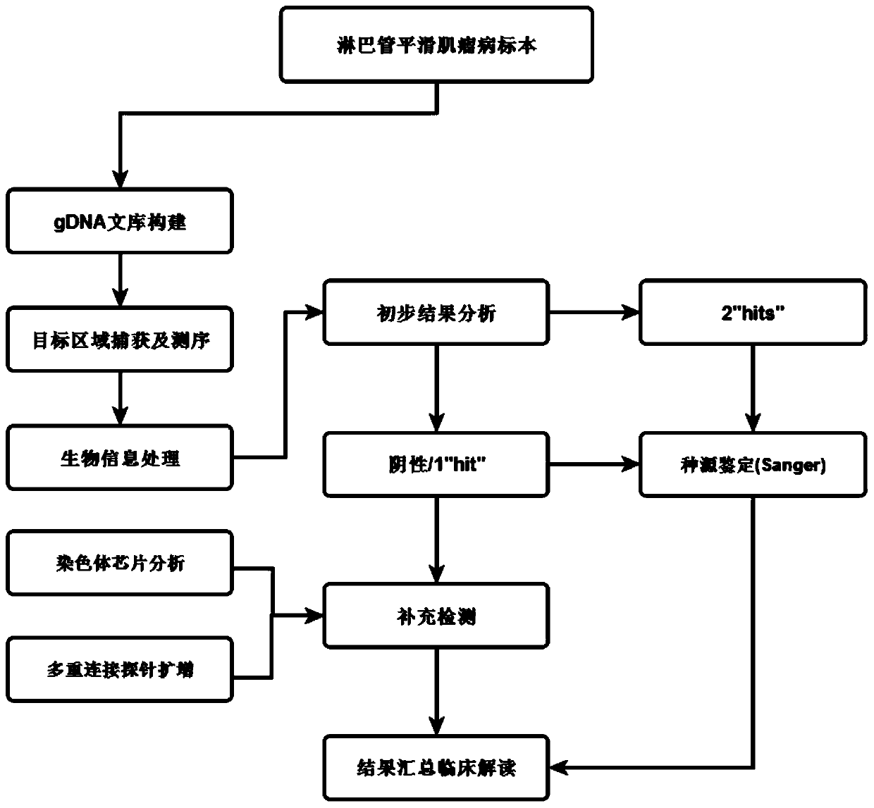
[0045] as per figure 1 In the multi-method combined detection scheme for lymphangioleiomyomatosis shown, the following detections are performed on lymphangioleiomyomatosis specimens.

[0046] 1. Using probes for liquid phase capture targeted sequencing (Target Capture Sequencing, NGS);

[0047] 1. Design Panel

[0048] The shingled design was performed on the entire exon coding regions of TSC1 and TSC2 genes that are highly related to LAM, and the probes of TSC1 and TSC2 genes were designed as follows: figure 1 As indicated, to ensure at least 2-fold coverage of the target regions of the two genes, each probe is 100 bp long.

[0049] At the same time, refer to COSMIC, TCGA and other databases, combined with the latest NCCN guidelines / consensus, screen the proto-cancer and tumor suppressor genes that are closely related to the occurrence and development of solid tumors, and design probes according to conventional methods, that is, the probes are connected at the ends. Perfor...

Embodiment 2

[0172] The method of Example 1 was used to carry out the LAM joint detection research.

[0173] 1. Comparison of single detection method and joint detection method.

[0174] In this study, a total of 61 patients with LAM were enrolled, and a single detection method and the method in Example 1 were used for detection at the same time.

[0175] A single detection method is a method that only uses NGS targeted sequencing.

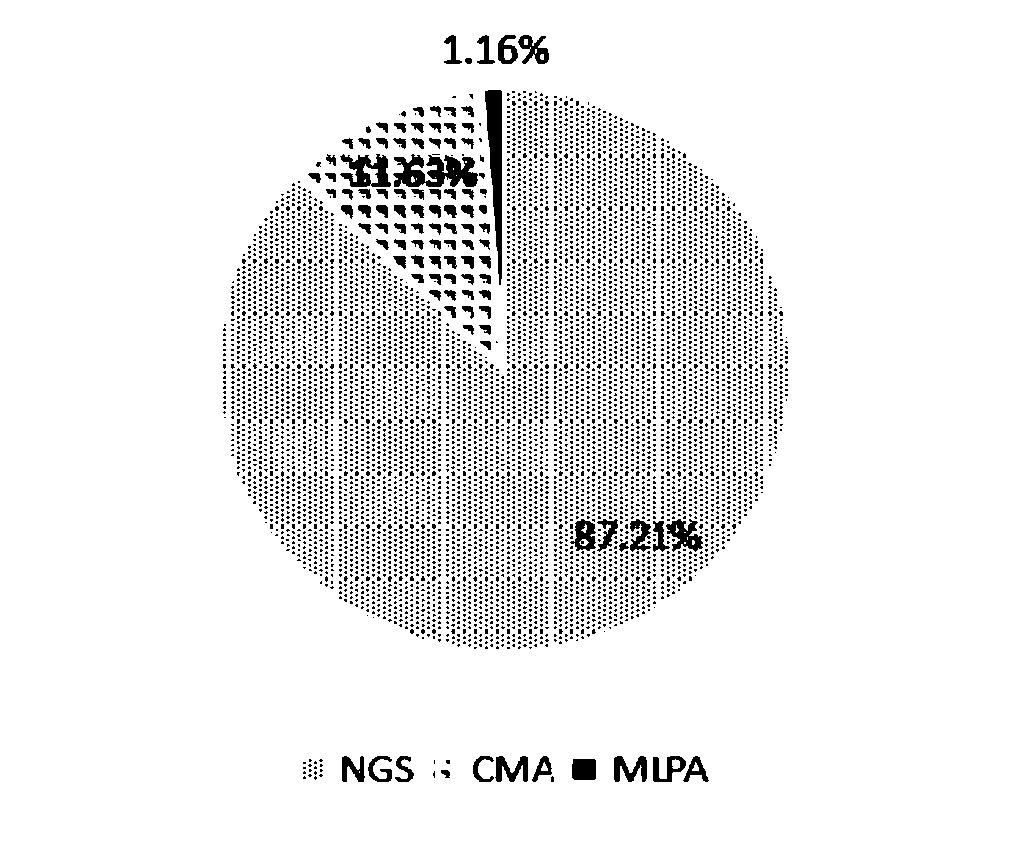
[0176] The result is as Figure 3-4 shown, where image 3 It is the detection ratio of variant sites using different detection methods, where NGS means liquid phase capture targeted sequencing, chromosome microarray analysis, and MLPA means multiple ligation probe amplification detection. The above results show that through multi-method joint detection, SNV, INDEL, CNV, LOH and other variant types can be fully output for LAM patient samples. If a single NGS detection method is used, 12.8% of the variants will be lost

[0177] Using a single detection metho...

Embodiment 3

[0185] An example of LAM joint detection is performed using the method of Embodiment 1.

[0186] 1. Sample source

[0187] The sample came from a fixed tissue specimen from the Respiratory Medicine Department of a tertiary hospital in Guangzhou, and the clinical diagnosis was S-LAM.

[0188] 2. Detection method and result

[0189] 1. Using probes for liquid phase capture targeted sequencing (Target Capture Sequencing, NGS)

[0190] Sequencing results showed that the sample had a single site mutation, TSC2: NM_000548.4:c.3412C>T(p.Arg1138*), and the mutation frequency was 50.9%.

[0191] 2. Chromosome microarray analysis and multiple junction probe amplification

[0192] According to the method of Example 1, the chromosome chip analysis method was used for supplementary detection, and the results showed that the patient found a loss of heterozygosity (LOH) mutation in the TSC2 gene: arr 16p13.3p11.2(83886_30809063)x2mos hmz, the variation size is 30.73Mb, its LOH fragment i...


PUM


Abstract

The invention relates to a lymphangiomyoma joint detection method and an application thereof, and belongs to the technical field of gene detection. The method comprises carrying out liquid phase capture targeting sequencing, designing Panel for TSC1 and TSC2 gene whole exon coding regions highly related to LAM and mutant genes closely related to solid tumors, constructing a gDNA library, carryingout hybrid capture, sequencing on a machine, carrying out sorting by analyzing the sequencing data through bioinformatics processing, and if the TSC1 and TSC2 genes are negative, carrying out supplementary detection by adopting a chromosome chip analysis method, if the single strike site is detected, performing supplementary detection by adopting multiplex ligation-dependent probe amplification, and if the sites have no clear sources of the somatic mutation and the embryonic system mutation, carrying out supplementary detection by adopting Sanger detection. The method improves the positive variation detection rate of LAM patients.

Description

technical field [0001] The invention relates to the technical field of gene detection, in particular to a combined detection method for lymphangioleiomyomatosis and its application. Background technique [0002] Lymphangioleio-myomatosis (LAM) is a rare multisystem neoplastic disease, almost all of which occur in women, and is characterized by chronic progressive diffuse thin-walled cystic lesions in both lungs. Patients with LAM have mild early symptoms, usually with gradually worsening dyspnea and recurrent pneumothorax and chylothorax. About 60-70% of patients will develop pneumothorax at some stage, and about 30% of patients will develop chylothorax. Common extrapulmonary manifestations include renal angiomyolipoma, retroperitoneal lymphatic involvement, and pelvic lymph node involvement. [0003] LAM can be divided into sporadic LAM (S-LAM) and tuberous sclerosis-associated LAM (TSC-LAM). The pathogenesis of LAM is still not very clear. At present, it is believed tha...

Claims


Application Information

Patent Timeline
IPC IPC(8): C12Q1/6858C12Q1/6886
CPCC12Q1/6858C12Q1/6886C12Q2600/156C12Q2600/16C12Q2535/122C12Q2537/143C12Q1/6869C12Q1/6883
Inventor 欧小华刘菲菲孙明明胡昌明邓俊豪于世辉赵薇薇
Owner GUANGZHOU KINGMED DIAGNOSTICS GRP CO LTD
Learn More