Methods for in vivo expansion of cd8+ t cells and prevention or treatment of gvhd
A technology of cells and therapeutic agents, applied in the field of in vivo expansion of CD8+ T cells to prevent or treat GVHD
- Summary
- Abstract
- Description
- Claims
- Application Information
AI Technical Summary
Problems solved by technology
Method used
Image
Examples
Embodiment 1
[0129] Example 1: Donor CD4 + Effects of T cell depletion on GVHD prevention and GVL retention
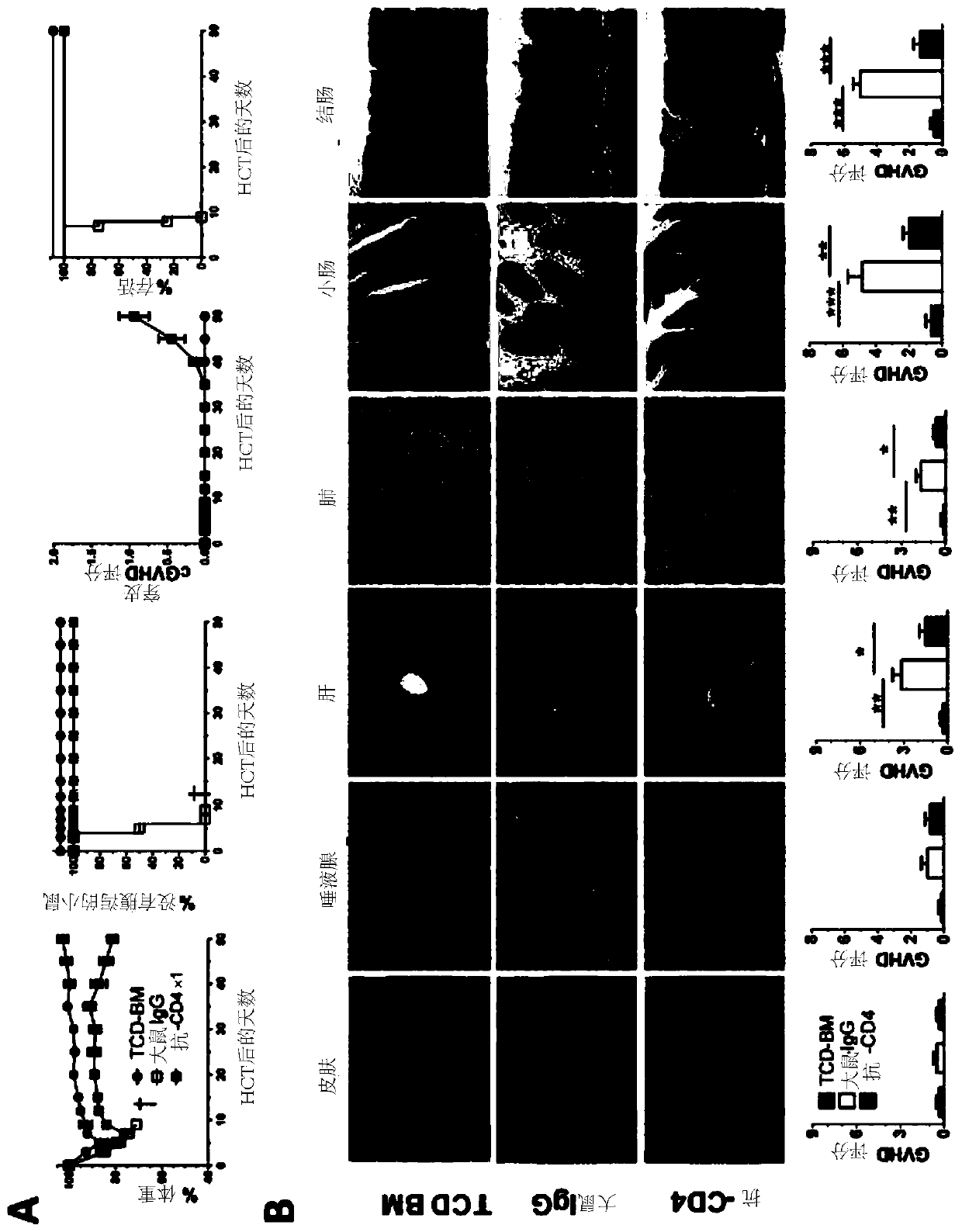
[0130] This example shows that donor CD4 after HCT + Immediate transient depletion of T cells preserves strong GVL effects while effectively preventing both acute and chronic GVHD in multiple models.
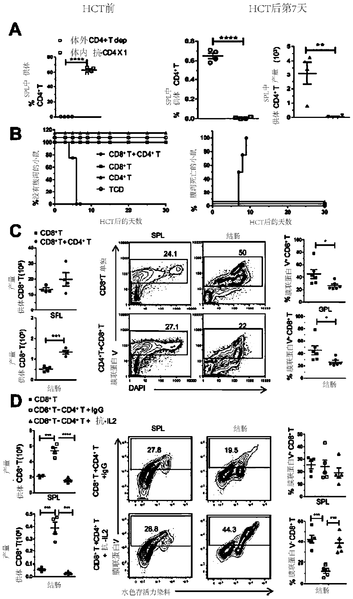
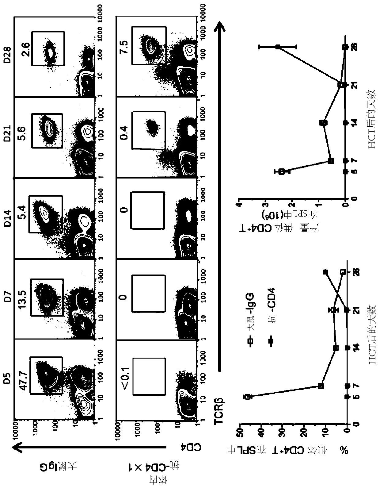
[0131] A previous study showed that sorted CD8 from C57BL / 6 donors + T cells did not induce acute GVHD, but did induce chronic GVHD in lethally irradiated BALB / c recipients, as shown by histopathology of salivary glands, the prototypical target organ of chronic GVHD. Anti-CD4 mAbs against CD4 on days 15 and 30 + Treatment of T cell depletion can prevent the development of chronic GVHD, as shown by the prevention of tissue damage in all GVHD target tissues, especially in the salivary glands (11). Disclosed herein is 1) Donor CD4 in recipient spleen 7 days after passing HCT + Percentage and yield judgment of T, and CD4 + In vivo administration of anti-CD4 mAb on the day of ...
Embodiment 2
[0141] Example 2: Donor CD4 + Effects of T cell depletion on IFN-γ and IL-2
[0142] This example demonstrates that donor CD4 immediately after HCT + Depletion of T cells increased serum IFN-γ but decreased serum IL-2 concentrations.
[0143] In experiments in C57BL / 6 donors and BALB / c recipients, in vivo donor CD4 immediately after HCT was explored + How T cell depletion prevents acute GVHD while preserving GVL effects. High serum levels of IFN-[gamma] and TNF-[alpha] have been associated with acute GVHD (41). Contrary to expectations, donor CD4 + Depletion of T cells increased serum IFN-γ concentrations by approximately 3-fold (p Figure 11 A). Elevated serum levels of IFN-γ are attributed to donor CD8 in lymphoid tissue + Expansion of T cells because of IFN-γ in the spleen of anti-CD4-treated recipients + CD8 + The number of T cells was approximately 3-fold higher than in rat IgG-treated recipients (p+ IFN-γ in T cells + The percentage of cells is similar ( F...
Embodiment 3
[0144] Example 3: Donor CD4 + Depletion of T cells affects donor CD8 in lymphoid tissue + The effect of T cell number
[0145] This example demonstrates that donor CD4 immediately after HCT + Depletion of T cells increases donor CD8 in lymphoid tissues + The number of T cells.
[0146] Next, the in vivo CD4 + T cell depletion on donor CD8 + Effects on T cell expansion and tissue distribution. H-2K in spleen and MLN of anti-CD4 treated recipients at day 5 after HCT b+ donor CD8 + The number of T cells was lower than that of rat IgG-treated recipients (P Figure 11 C). Donor CD8 in the spleen, PLN, and MLN of anti-CD4-treated recipients 7–10 days after HCT + The number of T cells was approximately 3-fold higher than in control IgG-treated recipients (p Figure 11 C). By day 28, donor CD8 + T cells re-expanded in the lymphoid tissue of anti-CD4 treated recipients but not in IgG treated recipients, and IgG treated recipients showed severe lymphopenia ( Figure 1...
PUM
 Abstract
Description
Claims
Application Information
 - R&D
- Intellectual Property
- Life Sciences
- Materials
- Tech Scout
- Unparalleled Data Quality
- Higher Quality Content
- 60% Fewer Hallucinations
Browse by: Latest US Patents, China's latest patents, Technical Efficacy Thesaurus, Application Domain, Technology Topic, Popular Technical Reports.
© 2025 PatSnap. All rights reserved.Legal|Privacy policy|Modern Slavery Act Transparency Statement|Sitemap|About US| Contact US: help@patsnap.com


