Medical image processing method and device, storage medium and computer equipment

A technology of medical images and processing methods, applied in the medical field, can solve the problems of being prone to errors, aggravating the risk of misdiagnosis, and low efficiency of manual observation and analysis

Pending Publication Date: 2020-04-10
SHANGHAI UNITED IMAGING INTELLIGENT MEDICAL TECH CO LTD
View PDF4 Cites 2 Cited by
  • Summary
  • Abstract
  • Description
  • Claims
  • Application Information

AI Technical Summary

Problems solved by technology

[0004] However, the efficiency of manual observation and analysis is low and error-prone, which will increase the risk of mi...

Method used


Image

  • Medical image processing method and device, storage medium and computer equipment
  • Medical image processing method and device, storage medium and computer equipment
  • Medical image processing method and device, storage medium and computer equipment

Examples


Embodiment Construction

[0030] In order to make the purpose, technical solution and advantages of the present application clearer, the present application will be further described in detail below in conjunction with the accompanying drawings and embodiments. It should be understood that the specific embodiments described here are only used to explain the present application, and are not intended to limit the present application.

[0031] In one embodiment, such as figure 1 As shown, a medical image processing method is provided, and the method is applied to a processor capable of medical image processing as an example for explanation. The method mainly includes the following steps:

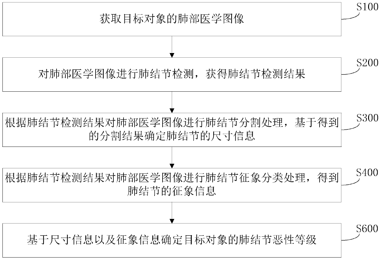
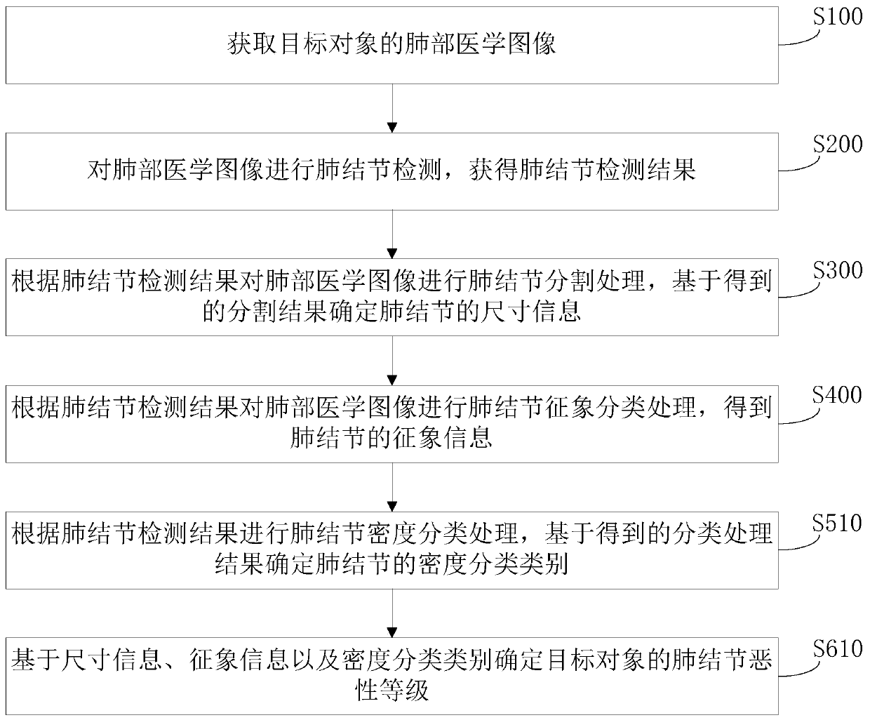
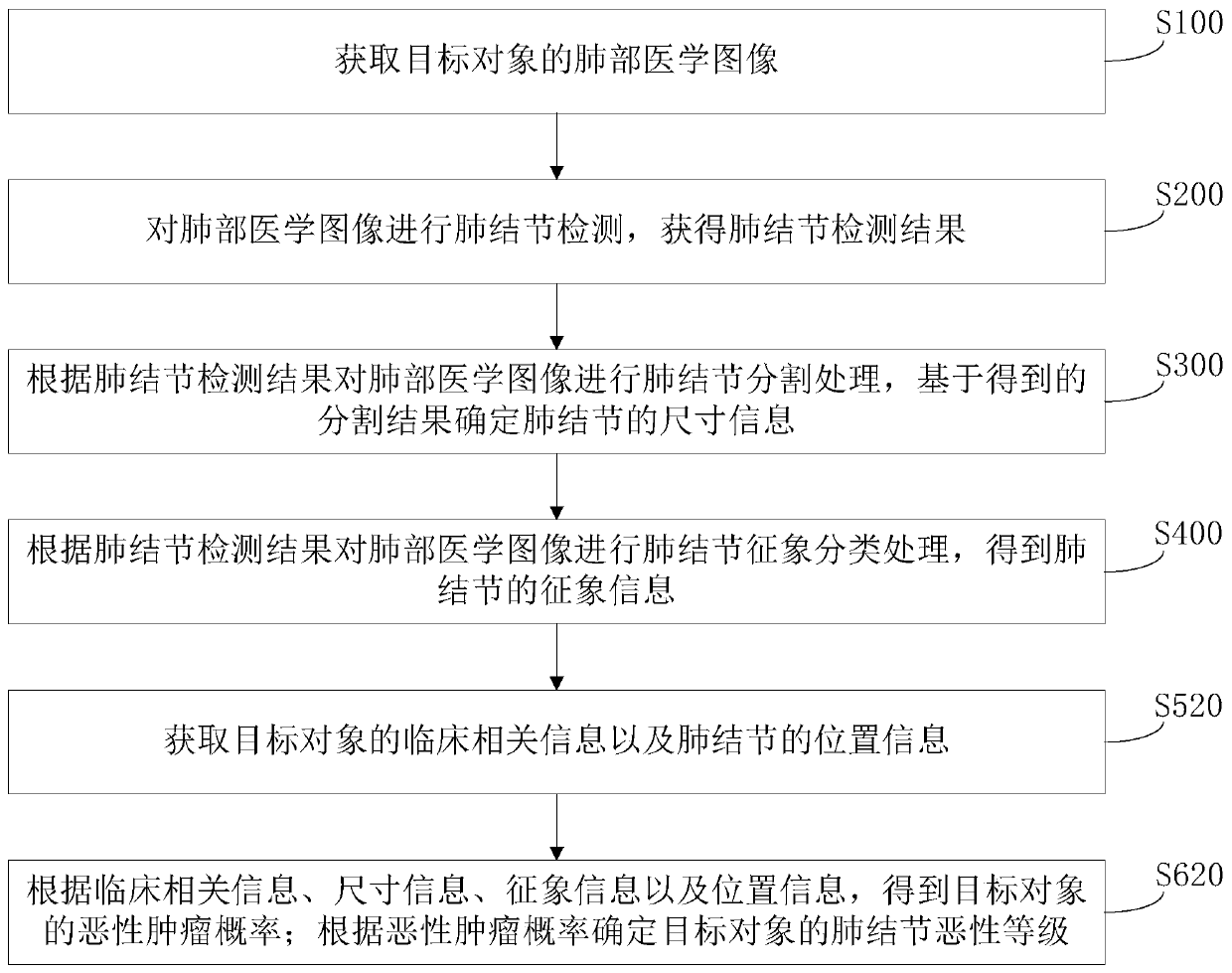
[0032] Step S100, acquiring a lung medical image of a target subject.

[0033] Specifically, the processor may perform image reconstruction and correction on the scanning data collected by the medical scanning device, so as to obtain the lung medical image of the target object. Of course, the medical image can also be...


PUM


Abstract

The invention relates to a medical image processing method and device, a storage medium and computer equipment. According to the method provided by the invention, after the lung medical image is acquired, pulmonary nodule segmentation processing and pulmonary nodule symptom classification processing can be automatically performed, then the malignant grade information of the pulmonary nodule is automatically determined according to the pulmonary nodule size information and symptom information obtained through processing, and compared with a manual observation method, the processing efficiency of the method provided by the invention is higher; besides, the method is carried out by integrating multiple aspects of information such as underlying features (namely size information) and advanced semantic information (namely symptom information), the accurate pulmonary nodule malignancy level can be obtained, later-period further diagnosis and definite diagnosis can be effectively supported, and the later-period misdiagnosis risk is remarkably reduced.

Description

technical field [0001] The present application relates to the field of medical technology, in particular to a medical image processing method, device, storage medium and computer equipment. Background technique [0002] Lung cancer is a malignant tumor with the highest incidence rate, the fastest-growing mortality rate, and the greatest threat to human health and life. The early manifestation of lung cancer is in the form of nodules. The earlier the discovery, the earlier the treatment, and the lower the chance of the target object suffering from lung cancer. Therefore, the detection of the malignant grade of lung nodules is of great significance to reduce the mortality of lung cancer. [0003] In the prior art, computed tomography (CT) is usually used for lung cancer screening, and doctors observe and analyze the obtained medical images to determine the benign and malignant development status of pulmonary nodules. [0004] However, the efficiency of manual observation and ...

Claims


Application Information

Patent Timeline
IPC IPC(8): G06T7/00G06T7/10G06T7/62
CPCG06T7/0012G06T7/10G06T7/62G06T2207/10081G06T2207/30064
Inventor 邵影高耀宗
Owner SHANGHAI UNITED IMAGING INTELLIGENT MEDICAL TECH CO LTD
Why Eureka
Try Eureka