Brain tumor segmentation method based on u-net network and multi-view fusion

A brain tumor and multi-view technology, applied in the field of 3D medical image segmentation, can solve the problems of V-Net's large amount of computation and the inability to restore image resolution well, so as to increase the receptive field of convolution kernel and reduce the difficulty of computation , the effect of high segmentation accuracy

Active Publication Date: 2022-02-15
TIANJIN UNIV
View PDF8 Cites 0 Cited by
  • Summary
  • Abstract
  • Description
  • Claims
  • Application Information

AI Technical Summary

Problems solved by technology

[0004] Aiming at the problem of 3D medical image segmentation, the present invention provides a method for 3D brain tumor MRI image segmentation based on cascaded anisotropic hole convolution network and multi-view fusion
It can effectively solve the problem that the FCN structure cannot restore the image resolution well, and at the same time solve the problem of the correlation information between the U-Net structure slices and the large amount of calculation of the V-Net; the segmentation result has high accuracy, and the technical solution is as follows:

Method used


Image

  • Brain tumor segmentation method based on u-net network and multi-view fusion
  • Brain tumor segmentation method based on u-net network and multi-view fusion
  • Brain tumor segmentation method based on u-net network and multi-view fusion

Examples


Embodiment Construction

[0028] In order to make the technical solution of the present invention clearer, the present invention will be further elaborated below in conjunction with the accompanying drawings. The present invention is concretely realized according to the following steps:

[0029] The first step is to build a dataset and perform data preprocessing:

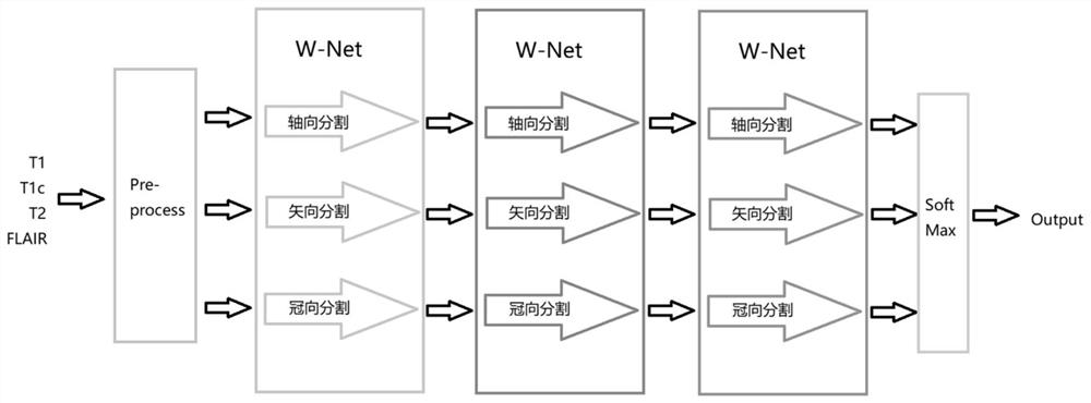
[0030] The present invention uses the published BraTS 2017 (Brain Tumor Segmentation 2017) data set, the data set consists of 210 high-glial tumor (HGG) cases and 75 low-glial tumor (LGG) cases, a total of 285 cases, the data set According to the ratio of 4:1 between the training set and the test set, 20% of the HGG and LGG cases are selected as the test set; each case contains MRI images of four modalities: T1, T1c, T2 and FLAIR. The size of each MRI image is 240*240*155. Due to the limitation of GPU memory and the increase of the training volume of the region of interest and the reduction of false positive points, the images of the traini...


PUM


Abstract

The present invention relates to a brain tumor segmentation method based on U-Net network and multi-view fusion, comprising the following steps: the first step is to preprocess the MRI images of brain tumors along the axial, sagittal and coronal directions Cut it into pieces suitable for the cascaded anisotropic atrous convolution U-Net network, and divide the training set and test set; the second step is to use the Tensorflow framework to construct the cascaded anisotropic atrous convolution U-Net network, cascading each The main structure of the anisotropic atrous convolution U-Net network is a three-level cascade: the network W-Net for segmenting the whole brain tumor, the T-Net for segmenting the tumor nucleus, and the network E-Net for segmenting the enhanced tumor , the three-level network is connected by cascading; the fourth step is to test the model.

Description

technical field [0001] The invention belongs to the field of 3D medical image segmentation, and relates to a 3D brain tumor nuclear magnetic resonance image segmentation method with cascaded anisotropic hole convolution U-Net network and multi-view fusion. Background technique [0002] Brain tumor segmentation is the separation of tumor regions from healthy tissue regions in brain medical images of patients with brain tumors. Among brain tumors, glial tumors are the most common tumors, which can be divided into high-glial tumors (HGG) and low-glial tumors (LGG) according to the aggressiveness and histological heterogeneity of tumor cells. The tumor area can be subdivided into tumor nucleus, edema, enhancing tumor, non-enhancing tumor and necrosis. Modern MRI images can effectively distinguish the above regions: T1, T1c, T2 and FLAIR four modalities respectively Areas of tumor nucleus, areas of enhancing tumor and necrosis, edema, and the entire tumor area are emphasized. D...

Claims


Application Information

Patent Timeline
Patent Type & Authority Patents(China)
IPC IPC(8): G06V10/764G06V10/774G06K9/62G06N3/04G06N3/08
CPCG06T7/11G06N3/084G06T2207/10088G06T2207/20081G06T2207/20084G06T2207/30096G06T2207/30016G06N3/045G06F18/24
Inventor 褚晶辉黄凯隆吕卫
Owner TIANJIN UNIV
Why Eureka
Try Eureka