
Single-channel fetal heart rate monitoring method based on search tree

A fetal heart rate, single-channel technology, applied in the measurement of pulse rate/heart rate, diagnostic recording/measurement, medical science, etc., can solve problems such as poor separation

Active Publication Date: 2020-04-24
SUN YAT SEN UNIV
View PDF9 Cites 0 Cited by
  • Summary
  • Abstract
  • Description
  • Claims
  • Application Information

AI Technical Summary

Problems solved by technology

Although the noise is excluded by the similarity of the QRS wave, for the Ni-FECG signal of the fetus, since the noise from the mother is similar to the waveform of the QRS wave of the fetus, it is very difficult to separate

Method used


Image

  • Single-channel fetal heart rate monitoring method based on search tree
  • Single-channel fetal heart rate monitoring method based on search tree
  • Single-channel fetal heart rate monitoring method based on search tree

Examples


Embodiment 1

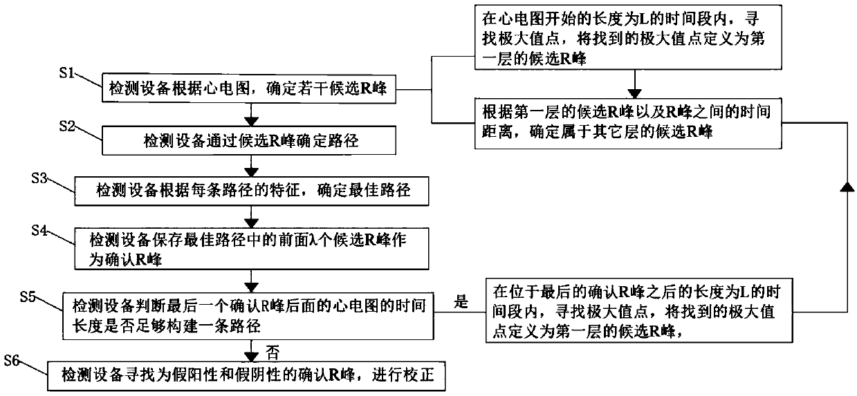
[0079] like figure 1 As shown, a method for realizing single-channel fetal heart rate monitoring based on search tree comprises the following steps:

[0080] The monitoring device finds the best path based on changes in the ECG, including:

[0081] Step S1: the monitoring device determines several candidate R peaks according to the electrocardiogram;

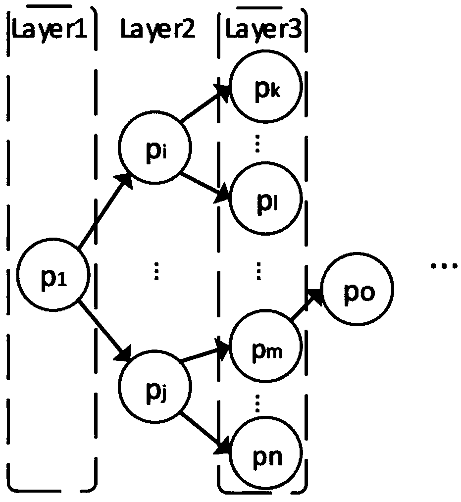
[0082] Step S2: The monitoring device determines the path through the candidate R peak;

[0083] Step S3: The monitoring device determines the best path of the QRS wave according to the characteristics of each path.

[0084] In step S1, the following steps are included:

[0085] S11. The monitoring device searches for a maximum value point within a time period of length L from the beginning of the electrocardiogram, and defines the found maximum value point as a candidate R peak of the first layer;

[0086] S12. According to the candidate R peaks of the first layer and the time distance between the R peaks, determine the ca...

Embodiment 2

[0114] Different from Embodiment 1, this embodiment also includes the following steps after step S3:

[0115] Step S4, the monitoring device saves the first λ candidate R peaks in the best path as confirmed R peaks;

[0116] Step S5, the monitoring device judges whether the time length of the electrocardiogram behind the last confirmed R peak is enough to construct a path (that is, judges whether the time of the last confirmed R peak is greater than or equal to the path limit length); In the period of time after the R peak whose length is L, search for a maximum point, define the found maximum point as a candidate R peak of the first layer, and execute step S12.

[0117] After step S5, step S6 is also included, the monitoring equipment searches for the confirmed R peaks of false positives and false negatives, and performs correction.

[0118] When looking for all false negative and false positive points, judge the time distance RR between all confirmed R peaks and their adjac...

Embodiment 3

[0122] In order to better explain step S5, the following example is cited in this embodiment. In step S3, the time points of candidate R peaks of each layer that have obtained the best path are 0.2s, 0.8s, 1.4s, 2.0s, 2.6s , since the λ value is 2, only the first two candidate R-peaks, namely the candidate R-peaks at time points 0.2s and 0.8s, are selected as the confirmation R-peaks. However, since the last time point for confirming the R peak is 0.8s, and the time span of the ECG is 30s, that is, there are still 29.2s in the following time, the time is obviously enough, which is much longer than the path limit length. For this reason, it is necessary to search for the R peak within a period of time after 0.8s in the electrocardiogram, perform a new round of optimal path selection, and search for more confirmed R peaks.


PUM


Abstract

The invention discloses a single-channel fetal heart rate monitoring method based on a search tree. The single-channel fetal heart rate monitoring method comprises the following steps: abdomen electrocardiogram pretreatment: cleaning of high-frequency noise and baseline drift of an electrocardiogram by monitoring equipment; and searching of an optimal path according to the similarity of the electrocardiogram by the monitoring equipment, wherein the steps comprise that S1, the monitoring equipment determines a plurality of candidate R peaks within a period of time according to the electrocardiograms within the period of time, S2, the monitoring equipment determines a path through the candidate R peak, and S3, the monitoring equipment determines the optimal path according to the characteristics of each path. According to the invention, with the method, a large amount of data can be reserved, so that the problem that the QRS wave, similar to the QRS wave of the fetus, of the mother is defined as the real QRS wave of the fetus according to the principle of similarity in the early stage of data processing while the real QRS waves of the fetus are excluded can be avoided, and the real QRS wave group can be found by comparing the similarity degrees of the QRS waves in different paths after the data are screened through the time distance between the QRS waves.

Description

technical field [0001] The invention relates to the field of fetal heart rate detection, and further provides a search tree-based single-channel fetal heart rate monitoring method with high accuracy. Background technique [0002] Fetal heart rate (FHR) is an important indicator for monitoring fetal health during pregnancy. Non-invasive fetal electrocardiogram (Ni-FECG) is a method of placing electrodes on the abdominal surface to obtain fetal electrocardiogram signals, which plays an important role in fetal heart rate measurement. [0003] Congenital heart disease is one of the leading causes of death from birth defects and is an urgent research topic today. Therefore, we need to develop an efficient technique to monitor fetal status during pregnancy and delivery. At present, the most widely used clinically is Doppler ultrasonography. However, it has not been conclusively proven that the use of ultrasound radiofrequency is completely safe for the fetus. Furthermore, the ...

Claims


Application Information

Patent Timeline
IPC IPC(8): A61B5/024A61B5/0472A61B5/366
CPCA61B5/02411A61B5/725A61B2503/02A61B5/366
Inventor 王国利钟伟郭雪梅
Owner SUN YAT SEN UNIV
Learn More