A kind of method for separating and culturing vaginal epithelial cells

A technique for separating and culturing vaginal epithelial cells is applied in the field of separating and culturing vaginal epithelial cells, and can solve the problems of small vaginal tissue and difficulty in separating the epithelium with dispase.

Active Publication Date: 2021-05-04
NANJING DRUM TOWER HOSPITAL +1
View PDF8 Cites 0 Cited by
  • Summary
  • Abstract
  • Description
  • Claims
  • Application Information

AI Technical Summary

Problems solved by technology

However, the reality is that clinically available human vaginal tissue is small and fragmented, making dispase isolation of the epithelium difficult.

Method used


Image

  • A kind of method for separating and culturing vaginal epithelial cells
  • A kind of method for separating and culturing vaginal epithelial cells
  • A kind of method for separating and culturing vaginal epithelial cells

Examples


Embodiment 1

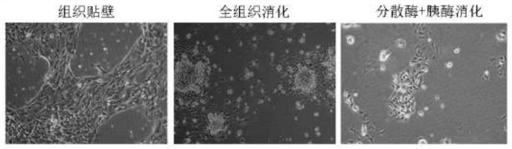
[0037] Cut up the tissue, digest it with 0.25g / 100ml% trypsin solution containing 0.02g / 100ml EDTA for 1 hour, then pass through a 70 micron cell sieve to collect the cells. Continue to add hyaluronidase (Sigma, No.H3506, 0.069g / 100ml), type I collagenase (Sigma, No.C0130, 0.049g / 100ml) and DNase (Roche, No.139-134P11, 0.012g / 100ml) mixture was digested for 30min, then passed through a 70 micron cell sieve, and the cells were collected. The cells collected twice were centrifuged (800g), washed twice with culture medium, and spread on Matrigel (BD, 356234) pre-coated cell culture plates to obtain epithelial cells with higher purity.

Embodiment 2

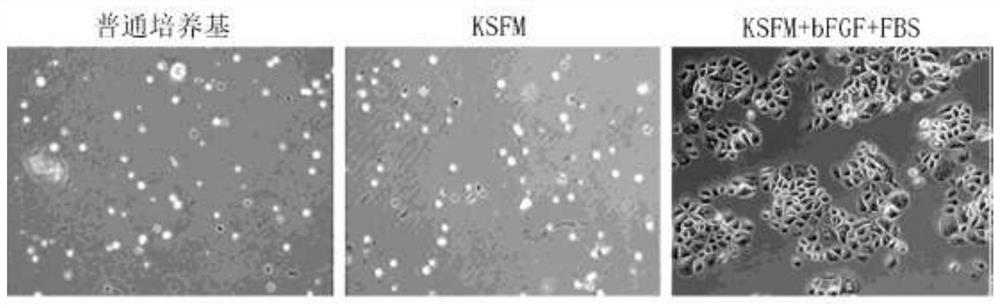
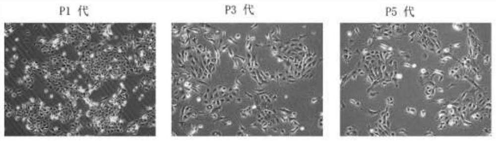
[0044]We evenly spread the obtained epithelial cells in 3 wells of Matrigel (BD, 356234) pre-coated cell culture plate. The cells in the three wells were added to ordinary medium (DF12 medium), KSFM medium and our improved KSFM medium. Such as figure 2 As shown, common culture medium and independent KSFM culture cannot maintain the growth of vaginal epithelial cells, but when we add 2% FBS and 5ng / ml bFGF, the growth and expansion of vaginal epithelial cells can be maintained ( figure 2 and image 3 ). After the epithelial cells were passed to the fifth generation, the surface markers were identified by immunofluorescence, and the results showed that the cultured cells were mainly epithelial cells (CK+), mixed with a very small amount of mesenchymal cells (vimentin+), and no fibroblasts ( Hsp47+) and vascular endothelial cells (CD34+) ( Figure 4 ).


PUM


Abstract

The invention discloses a method for separating and culturing vaginal epithelial cells. A method for separating and culturing vaginal epithelial cells, comprising the following steps: chopping up the tissue, first digesting it with digestive solution A, then passing it through a 70-micron cell sieve, and collecting the cells; adding the remaining tissue to the mixed digestive solution for digestion, and then passing it through a 70-micron cell sieve , to collect the cells; the cells collected twice were centrifuged, washed several times with the medium, and spread on Matrigel pre-coated cell culture plates to obtain vaginal epithelial cells. In the present invention, for the first time, vaginal epithelial cells are obtained by secondary digestion and separation of vaginal tissue with EDTA-containing trypsin and mixed digestive juice. The growth and expansion of vaginal epithelial cells can be maintained by culturing vaginal epithelial cells in optimized KSFM medium.

Description

technical field [0001] The invention belongs to the technical field of cell culture, and relates to a method for separating and culturing vaginal epithelial cells. Background technique [0002] Patients with congenital absence of vagina or vaginal hypoplasia, absent or narrow vagina after pelvic exenteration often need vaginal reconstruction. The standardized preparation of tissue-engineered vaginal seed cells is a prerequisite for functional vaginal reconstruction. However, autologous vaginal epithelial cells are difficult to passage, easy to mutate, and difficult to standardize and popularize. Therefore, it is necessary to further explore the isolation, culture and expansion technology of the cells. [0003] At present, vaginal epithelial cells are mostly digested with dispase overnight, and after the epithelium is peeled off, they are obtained by further digestion with trypsin. Then the isolated epithelial cells were plated and cultured with epithelial-specific medium K...

Claims


Application Information

Patent Timeline
Patent Type & Authority Patents(China)
IPC IPC(8): C12N5/071
CPCC12N5/0625C12N5/0682C12N2501/115C12N2509/00C12N2533/90
Inventor 赵光锋胡娅莉戴建武
Owner NANJING DRUM TOWER HOSPITAL
Why Eureka
Try Eureka