
Magnetic resonance positive contrast imaging method and device

An imaging method and imaging device technology, applied in magnetic resonance measurement, measurement using nuclear magnetic resonance imaging system, medical imaging, etc., can solve the problem of slow convergence of conjugate gradient algorithm, difficulty in distinguishing tissue gaps and low signal-to-noise ratio areas, and inability to Accurate positioning and evaluation of magnetic resonance compatible devices to achieve the effect of improving image reconstruction speed and reducing reconstruction complexity

Active Publication Date: 2021-03-23
SHENZHEN INST OF ADVANCED TECH CHINESE ACAD OF SCI
View PDF13 Cites 0 Cited by
  • Summary
  • Abstract
  • Description
  • Claims
  • Application Information

AI Technical Summary

Problems solved by technology

MRI compatible devices for human intervention / implantation are generally made of metal. Since metals are easily magnetized in a magnetic field and will generate a local magnetic field, the MRI compatible device and its neighborhood will show negative contrast in the image. Contrast is indistinguishable from tissue voids and areas of low signal-to-noise ratio, preventing precise localization and evaluation of MR-compatible devices
[0003] In recent years, in order to accurately locate and evaluate MRI compatible devices, researchers have proposed a series of MRI positive contrast imaging techniques, using the conjugate gradient algorithm to iteratively solve the magnetic susceptibility map, but the conjugate gradient algorithm converges slowly, especially in the vicinity of The optimal solution requires multiple loop iterations, resulting in slower image reconstruction time

Method used


Image

  • Magnetic resonance positive contrast imaging method and device
  • Magnetic resonance positive contrast imaging method and device
  • Magnetic resonance positive contrast imaging method and device

Examples


no. 1 example

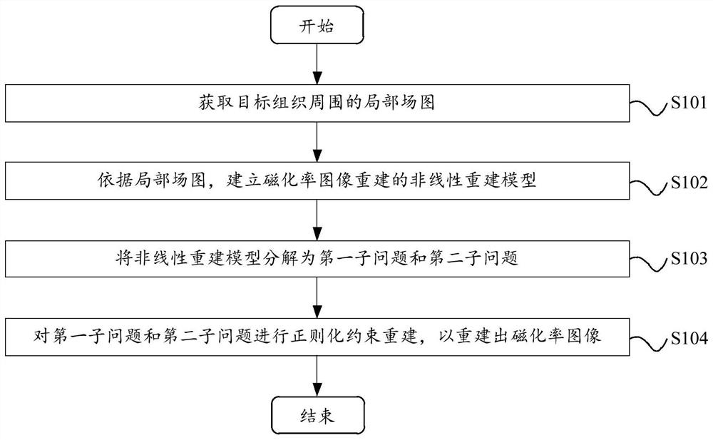
[0026] Please refer to figure 2 , figure 2 A flow chart of the magnetic resonance positive contrast imaging method provided by the embodiment of the present invention is shown. The magnetic resonance positive contrast imaging method includes the following steps:

[0027] Step S101, acquiring a local field map around the target tissue.

[0028] In the embodiment of the present invention, the local field map around the target tissue is the local magnetic field generated by the metal device in the target area to the surrounding tissue, and the target area is the biological tissue containing the metal device. Methods to obtain a local field map around the target tissue can be:

[0029] Firstly, the data acquisition of the imaging target is carried out by the magnetic resonance scanner, and the magnetic resonance (Magnetic Resonance, MR) signal with the echo readout gradient offset is obtained; then, the phase of the MR signal with the echo readout gradient offset is used Obt...

no. 2 example

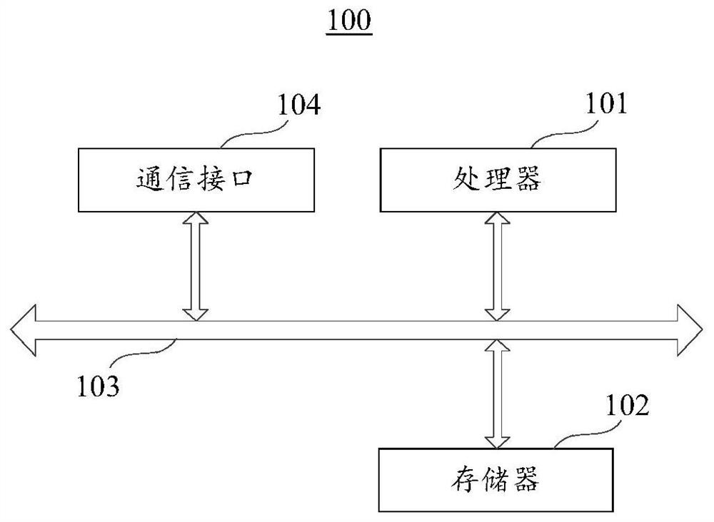
[0057] Please refer to Figure 5 , Figure 5 A schematic block diagram of a magnetic resonance positive contrast imaging apparatus 200 provided by an embodiment of the present invention is shown. The magnetic resonance positive contrast imaging device 200 includes a local field map acquisition module 201 , a model building module 202 , a model decomposition module 203 and an image reconstruction module 204 .

[0058] The local field map acquisition module 201 is configured to obtain a local field map around the target tissue.

[0059] The model establishing module 202 is configured to establish a nonlinear reconstruction model for magnetic susceptibility image reconstruction according to the local field diagram.

[0060] In the embodiment of the present invention, the model establishment module 202 is specifically used to establish a nonlinear reconstruction model for magnetic susceptibility image reconstruction according to the local field map Among them, ΔB represents th...


PUM


Abstract

Embodiments of the invention relate to the technical field of magnetic resonance imaging, and provide a magnetic resonance positive contrast imaging method and device. The method comprises: acquiringa local field map around a target tissue, collected by a magnetic resonance scanner; according to the local field map, establishing a nonlinear reconstruction model of magnetic susceptibility image reconstruction; decomposing the nonlinear reconstruction model into a first sub-problem and a second sub-problem; and performing regularized constraint reconstruction on the first sub-problem and the second sub-problem, so as to reconstruct a magnetic susceptibility image. Compared with the prior art, the embodiments of the invention decompose the complex reconstruction problem into the two sub-problems with lower complexity to solve, thereby reducing the reconstruction complexity and improving the image reconstruction speed.

Description

technical field [0001] Embodiments of the present invention relate to the technical field of magnetic resonance imaging, and in particular, to a magnetic resonance positive contrast imaging method and device. Background technique [0002] Magnetic Resonance Imaging (MRI) is a technology that uses the principle of magnetic resonance to perform tomographic imaging of the human body. Its advantages of no radiation, good soft tissue contrast, and arbitrary plane imaging make it a very popular clinical diagnostic tool. MRI compatible devices for human intervention / implantation are generally made of metal. Since metals are easily magnetized in a magnetic field and will generate a local magnetic field, the MRI compatible device and its neighborhood will show negative contrast in the image. Contrast is indistinguishable from tissue voids and areas of low signal-to-noise ratio, preventing precise localization and evaluation with MR-compatible devices. [0003] In recent years, in or...

Claims


Application Information

Patent Timeline
Patent Type & Authority Patents(China)
IPC IPC(8): G01R33/54A61B5/055
CPCA61B5/055A61B5/72A61B5/7203A61B2576/00G01R33/54
Inventor 王海峰梁栋郑海荣刘新程静史彩云陈汉威谢国喜苏适
Owner SHENZHEN INST OF ADVANCED TECH CHINESE ACAD OF SCI
Learn More