
Intensity self-calibration multi-channel X-ray imaging system and application method

An imaging system and X-ray technology, applied in the field of X-ray imaging, can solve the problems of difficult installation and adjustment, many channel scanning tasks, and long time-consuming, and achieve the effect of shortening the research and development cycle, improving the calibration efficiency, and strong adaptability

Active Publication Date: 2020-05-08
TONGJI UNIV
View PDF11 Cites 0 Cited by
  • Summary
  • Abstract
  • Description
  • Claims
  • Application Information

AI Technical Summary

Problems solved by technology

[0005] The purpose of the present invention is to provide an intensity self-calibration multi-channel X-ray imaging system and its application method in order to overcome the defects of difficult installation and adjustment, many channel scanning tasks and long time consumption in the prior art

Method used


Image

  • Intensity self-calibration multi-channel X-ray imaging system and application method
  • Intensity self-calibration multi-channel X-ray imaging system and application method
  • Intensity self-calibration multi-channel X-ray imaging system and application method

Examples


Embodiment 1

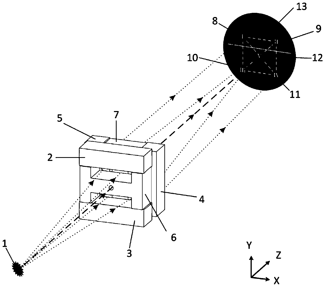
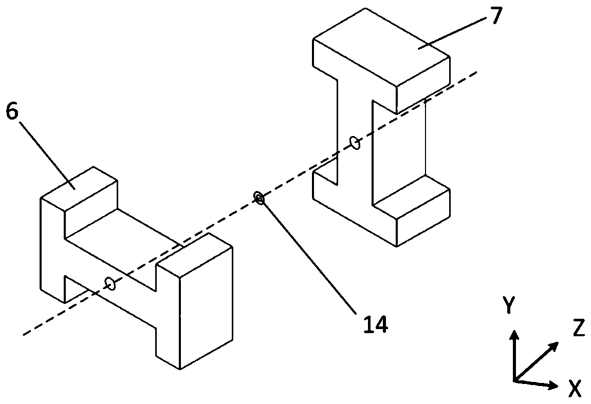
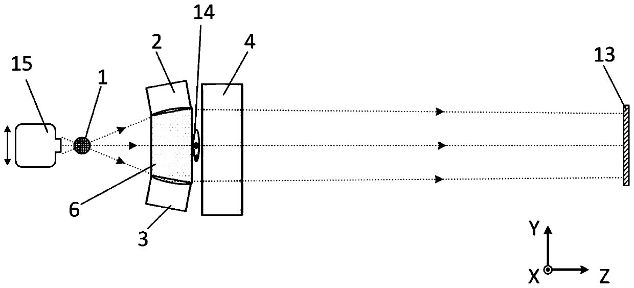
[0046] Select the first transverse cylindrical reflector 2, the second transverse cylindrical reflector 3, the first longitudinal cylindrical reflector 4 and the second longitudinal cylindrical reflector 5 with a radius of curvature of 50m, and the horizontal I-shaped optical cone The core 6 and the vertical I-shaped optical cone core 7 form a four-channel KB microscope. The initial structural parameters of the multi-channel X-ray imaging system are shown in Table 1:

[0047] Table 1 Initial structural parameters of the multi-channel X-ray imaging system

[0048]

[0049] The surfaces of the first transverse cylindrical reflector 2, the second transverse cylindrical reflector 3, the first longitudinal cylindrical reflector 4 and the second longitudinal cylindrical reflector 5 adopt double-layer X-ray film, and one layer is 30nm thickness The Pt film can be adapted to the energy points of 2.5, 6 and 8keV, and the single mirror reflectance is above 70%, which can meet the req...


PUM


Abstract

The invention relates to an intensity self-calibration multi-channel X-ray imaging system, which comprises a horizontal I-shaped optical cone core, a vertical I-shaped optical cone core, a pinhole plate and a large-format X-ray film camera for imaging, wherein transverse cylindrical reflectors are arranged at the top end and the bottom end of the horizontal I-shaped optical cone core, longitudinalcylindrical reflectors are arranged on the two sides of the vertical I-shaped optical cone core, the horizontal I-shaped optical cone core and the vertical I-shaped optical cone core are sequentiallyand orthogonally arranged in an attached mode, a small hole is formed in the center of each component, the small holes are coaxially aligned, the pinhole plate is located between the horizontal I-shaped optical cone core and the vertical I-shaped optical cone core, and the central axis of the pinhole plate is coaxial with the axis where the small holes are located. Compared with the prior art, the intensity self-calibration multi-channel X-ray imaging method has the advantages of being high in applicability, small in view field difference, capable of shortening the research and development period and the like.

Description

technical field [0001] The invention relates to the field of X-ray imaging, in particular to an intensity self-calibrating multi-channel X-ray imaging system and an application method. Background technique [0002] Pinhole cameras, X-ray microscopes, bent crystals, and coded imaging components play an important role in the diagnosis of laser inertial confinement fusion (ICF) in my country. Among them, as a linear space-invariant system, the pinhole camera has a relatively simple structure and can easily form a pinhole array, but the spatial resolution is affected by both geometric dimensions and diffraction effects. The spatial resolution of the aperture camera is between 20 μm and 40 μm. The disadvantages of pinhole cameras mainly lie in insufficient spatial resolution and light collection efficiency. The optical configurations of common X-ray microscopes mainly include KB type, KBA type, AKB type, and Wolter type. Among them, the Kirkpatrick-Baez (KB) X-ray microscope i...

Claims


Application Information

Patent Timeline
IPC IPC(8): G01N23/04
CPCG01N23/04
Inventor 徐捷穆宝忠王新
Owner TONGJI UNIV
Learn More