Structural illumination rapid three-dimensional color microscopic imaging method based on Hilbert transform
A technology of structured illumination and microscopic imaging, applied in the fields of microscopy, image analysis, image data processing, etc., can solve the problems of time-consuming storage and processing, large amount of data collection, etc., achieve image processing speed improvement, expand application scope, The effect of image acquisition reduction
- Summary
- Abstract
- Description
- Claims
- Application Information
AI Technical Summary
Problems solved by technology
Method used
Image
Examples
Embodiment 1
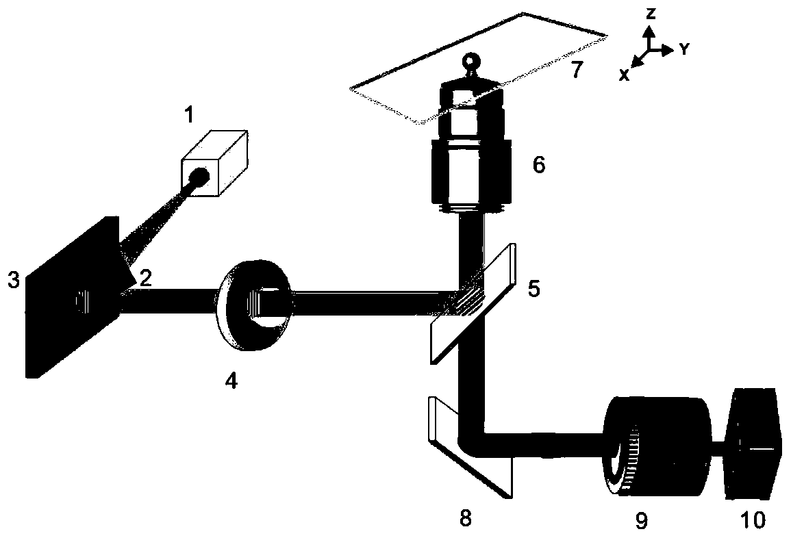
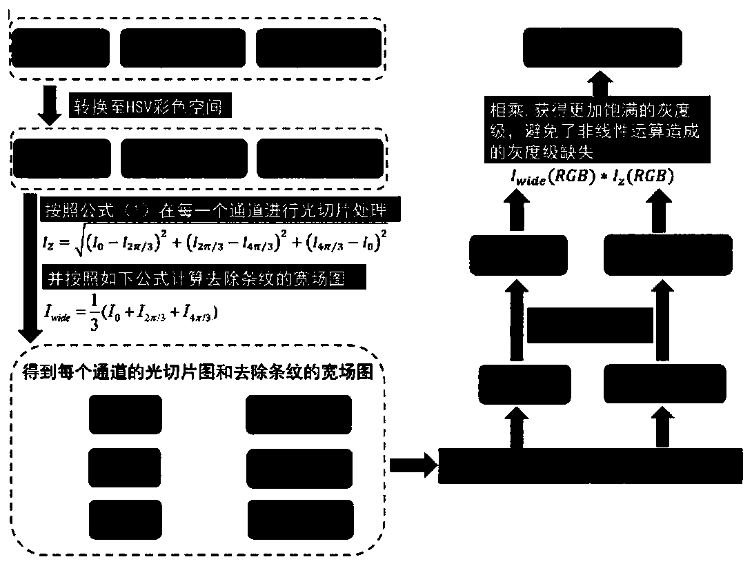
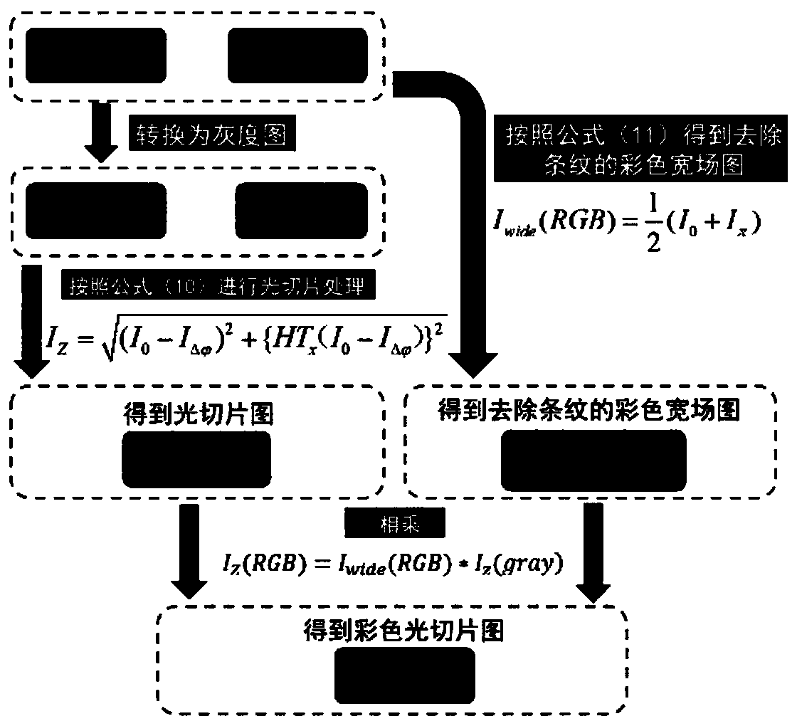
[0080] In order to verify the accuracy of the HT-COS method, pollen samples with autofluorescence (405nm excitation wavelength) were selected for experiments. Replace the 50:50 beamsplitter with a 425 nm long-pass dichroic mirror with a violet LED light source with a wavelength of 405 nm. At each focal plane of the sample, three original structured illumination images with a phase shift difference of 2π / 3 and two original structured illumination images with a phase shift difference of π were collected with an exposure time of 20ms, and the HSV-RMS algorithm and this Invented HT-COS algorithm for processing, the experimental results are as follows Figure 4 shown. Figure 4 (a) is the three-dimensional color light slice image processed by the HSV-RMS algorithm, Figure 4(b) is the result after processing by HT-COS algorithm. Obviously, there is no essential difference in the image restoration quality of the two algorithms, and the color restoration degree is basically the sa...
Embodiment 2
[0083] The original animal star sand in the ocean was imaged, and the original image was processed using the HSV-RMS algorithm and the HT-COS algorithm respectively. The results are as follows: Figure 5 shown. Figure 5 (a) is the maximum projection image of the star sand sample, taken with a 10X, NA0.45 objective lens, and stitched together 16 fields of view; Figure 5 (b-c) The field of view shown in the red box in (a) was shot with a 10X, NA0.45 objective lens, a total of 344 layers were shot, the layer spacing was 500nm, and the image size was 2048x2048 pixels; Figure 5 (b) is the original structured illumination image of the 64th layer of the sample; Figure 5 (c) A color wide-field image of the striped layer 64 of the sample; Figure 5 (d) is the maximum projection result reconstructed by the color SIM optical slice algorithm based on the HSV-RMS algorithm for all 344 layers of the field of view of the sample, and a total of 1032 original images were collected; Fig...
PUM
 Abstract
Description
Claims
Application Information
 - R&D
- Intellectual Property
- Life Sciences
- Materials
- Tech Scout
- Unparalleled Data Quality
- Higher Quality Content
- 60% Fewer Hallucinations
Browse by: Latest US Patents, China's latest patents, Technical Efficacy Thesaurus, Application Domain, Technology Topic, Popular Technical Reports.
© 2025 PatSnap. All rights reserved.Legal|Privacy policy|Modern Slavery Act Transparency Statement|Sitemap|About US| Contact US: help@patsnap.com


