A fast three-dimensional color microscopy imaging method based on structured illumination based on Hilbert transform

A structured lighting and microscopic imaging technology, which is applied in microscopes, image analysis, image data processing, etc., can solve the problems of large amount of time-consuming storage and processing and large amount of data collection, so as to improve the speed of image processing, expand the scope of application, The effect of reducing the amount of image acquisition

Active Publication Date: 2020-11-06
XI AN JIAOTONG UNIV
View PDF7 Cites 0 Cited by
  • Summary
  • Abstract
  • Description
  • Claims
  • Application Information

AI Technical Summary

Problems solved by technology

[0006] In order to solve the technical problems of the existing full-color 3D imaging method for size-level samples, which have a large amount of data collection and a large amount of time for storage and processing, the present invention provides a fast 3D color display with structured illumination based on Hilbert transform. Micro imaging method, compared with the existing color imaging algorithm based on HSV color space, in the imaging method provided by the present invention, the amount of image acquisition is reduced by 1 / 3, the image reconstruction speed is increased by about 28%, and the large-scale The high-resolution full-color three-dimensional imaging of the sample has laid the foundation, which will further expand the application range of SIM technology, and has broad application prospects in paleontology, zoology, material science and other fields

Method used


Image

  • A fast three-dimensional color microscopy imaging method based on structured illumination based on Hilbert transform
  • A fast three-dimensional color microscopy imaging method based on structured illumination based on Hilbert transform
  • A fast three-dimensional color microscopy imaging method based on structured illumination based on Hilbert transform

Examples


Embodiment 1

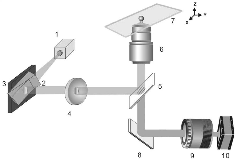
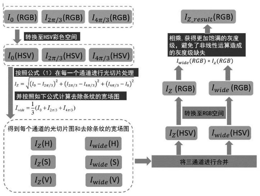
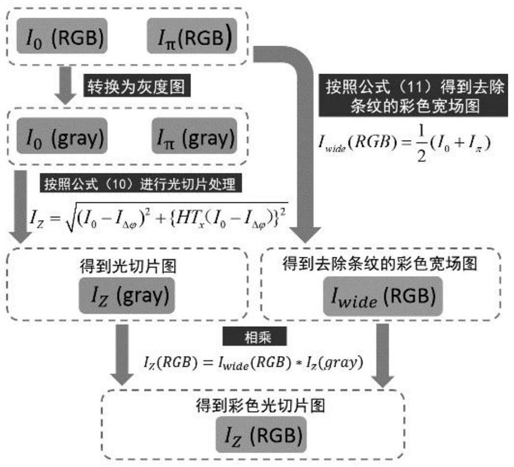
[0080] In order to verify the accuracy of the HT-COS method, pollen samples with autofluorescence (405nm excitation wavelength) were selected for experiments. Replace the 50:50 beamsplitter with a 425nm longpass dichroic mirror with a 405nm violet LED light source. At each focal plane of the sample, three original structured illumination images with a phase shift difference of 2π / 3 and two original structured illumination images with a phase shift difference of π were collected with an exposure time of 20ms, and the HSV-RMS algorithm and this Invented HT-COS algorithm for processing, the experimental results are as follows Figure 4 shown. Figure 4 (a) is the three-dimensional color light slice image processed by the HSV-RMS algorithm, Figure 4(b) is the result after processing by HT-COS algorithm. Obviously, there is no essential difference in the image restoration quality of the two algorithms, and the color restoration degree is basically the same. However, the "claw"...

Embodiment 2

[0083] The original animal star sand in the ocean was imaged, and the original image was processed using the HSV-RMS algorithm and the HT-COS algorithm respectively. The results are as follows: Figure 5 shown. Figure 5 (a) is the maximum projection image of the star sand sample, taken with a 10X, NA0.45 objective lens, and stitched together 16 fields of view; Figure 5 (b-c) The field of view shown in the red box in (a) was shot with a 10X, NA0.45 objective lens, a total of 344 layers were shot, the layer spacing was 500nm, and the image size was 2048x2048 pixels; Figure 5 (b) is the original structured illumination image of the 64th layer of the sample; Figure 5 (c) A color wide-field image of the striped layer 64 of the sample; Figure 5 (d) is the maximum projection result reconstructed by the color SIM optical slice algorithm based on the HSV-RMS algorithm for all 344 layers of the field of view of the sample, and a total of 1032 original images were collected; Fig...


PUM


Abstract

The invention relates to a structural illumination rapid three-dimensional color microscopic imaging method based on Hilbert transform, a full-color light slice image can be reconstructed only by acquiring two original images with any phase shift difference on each focal plane of a sample, the image acquisition amount is reduced by 1 / 3, and the image reconstruction time is reduced by about 28%.

Description

technical field [0001] The invention relates to a structured illumination microsystem, which can realize high-speed color three-dimensional tomographic imaging, and can be widely used in researches in the fields of biology, medicine, material science, microelectronics and the like. Background technique [0002] Color information is an important physical quantity to describe the characteristics of objects. The color tomography technology of 3D objects is an important means to obtain the surface morphological characteristics of objects, and it is also the basis for 3D digitization of real objects. The point-scanning microscopic imaging technology represented by the laser confocal scanning microscope has the ability of three-dimensional tomographic imaging. Combined with the multi-channel fusion technology, the color information of the fluorescent markers on the sample surface can be obtained, which further improves the contrast and resolution of the imaging. However, Due to th...

Claims


Application Information

Patent Timeline
Patent Type & Authority Patents(China)
IPC IPC(8): G02B21/06G02B21/36G06T7/90
CPCG02B21/06G02B21/365G02B21/367G06T7/90
Inventor 雷铭
Owner XI AN JIAOTONG UNIV
Why Eureka
Try Eureka