Image classification method and device, computer equipment and readable storage medium

A classification method and image technology, applied in the field of image processing, can solve the problems of low efficiency and accuracy in the lesion identification process, and achieve the effect of improving efficiency and accuracy

Pending Publication Date: 2020-05-15
SHANGHAI UNITED IMAGING INTELLIGENT MEDICAL TECH CO LTD
View PDF3 Cites 18 Cited by
  • Summary
  • Abstract
  • Description
  • Claims
  • Application Information

AI Technical Summary

Problems solved by technology

[0005] Based on this, it is necessary to provide an image classification method, device, computer equipment and readable storage medium for the problem that the efficiency and accuracy of the lesion identification process in the traditional technology are relatively low.

Method used


Image

  • Image classification method and device, computer equipment and readable storage medium
  • Image classification method and device, computer equipment and readable storage medium
  • Image classification method and device, computer equipment and readable storage medium

Examples


Embodiment Construction

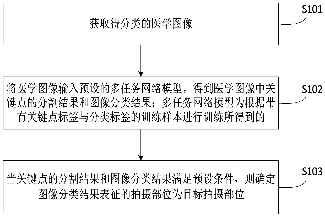
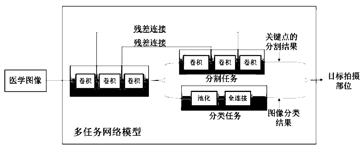
[0052] The image classification method provided by the embodiment of the present application can be applied to a process of classifying a photographed medical image to determine the photographing position of the medical image. The medical image may be an X-ray film, or a Computed Tomography (CT), a Nuclear Magnetic Resonance Imaging (MRI) or a Positron Emission Computed Tomography (PET), etc. . In the traditional technology, the part label of the medical image is usually entered by the scanning technician, and the radiologist identifies the lesions in the medical image according to the part label. However, the scanning technician may make mistakes and record the wrong part label. The efficiency and accuracy of the process of re-judging the scanning site and identifying the lesions are relatively low. The image classification method, apparatus, computer device, and readable storage medium provided by the embodiments of the present application are intended to solve the above-me...


PUM


Abstract

The invention relates to an image classification method and device, computer equipment and a readable storage medium. The method comprises the steps of obtaining a to-be-classified medical image; inputting the medical image into a preset multi-task network model to obtain a segmentation result and an image classification result of key points in the medical image; the multi-task network model beingobtained by training according to a training sample with a key point label and a classification label; and when the segmentation result of the key points and the image classification result meet a preset condition, determining the shooting part represented by the image classification result as a target shooting part. According to the method, the computer equipment determines the shooting part inthe medical image by adopting the multi-task network model, so that the accuracy of a target shooting part result can be improved, and the accuracy of a subsequent focus identification process is further improved; a radiologist does not need to make a confirmation process, and meanwhile, the efficiency of a focus recognition process is improved.

Description

technical field [0001] The present application relates to the technical field of image processing, and in particular, to an image classification method, apparatus, computer equipment and readable storage medium. Background technique [0002] X-rays (X-Rays) play an important role in the early detection of lung diseases, heart diseases, abdominal diseases and fractures due to their low price and better imaging results. Usually, after scanning the patient with medical equipment, the scanning technician submits the medical image to the corresponding radiologist for reading. [0003] Usually, when scanning a certain part of a patient, a scanning technician will input the scanned part label (for example, the current medical image is an abdominal image), and then the radiologist will identify the lesions in the medical image according to the part label. [0004] However, the traditional technology may record the wrong part label due to the mistake of the scanning technician, whic...

Claims


Application Information

Patent Timeline
Patent Type & Authority Applications(China)
IPC IPC(8): G06K9/46G06K9/62
CPCG06V10/462G06V2201/031G06F18/29G06F18/241
Inventor 詹恒泽梁凯轶周慧郑介志
Owner SHANGHAI UNITED IMAGING INTELLIGENT MEDICAL TECH CO LTD
Why Eureka
Try Eureka