
Rectal cancer pathology image classification method based on multi-channel collaborative capsule network

A pathology, multi-channel technology, applied in the direction of understanding medical/anatomical models, instruments, calculations, etc., can solve problems such as no rotation, loss of spatial information, etc., to improve generalization ability and overcome the effect of loss of spatial information

Active Publication Date: 2020-05-22
ZHEJIANG UNIV OF TECH
View PDF3 Cites 12 Cited by
  • Summary
  • Abstract
  • Description
  • Claims
  • Application Information

AI Technical Summary

Problems solved by technology

The present invention effectively overcomes the problems of traditional convolutional neural network technology such as loss of spatial information, lack of rotation and translation invariance in the training process, and uses feature fusion to improve the generalization ability of the model. The capsule layer allows collaborative cooperation between channels to accelerate Training, improving the accuracy of the capsule network in colon cancer pathology classification and reducing the training time of the model

Method used


Image

  • Rectal cancer pathology image classification method based on multi-channel collaborative capsule network
  • Rectal cancer pathology image classification method based on multi-channel collaborative capsule network
  • Rectal cancer pathology image classification method based on multi-channel collaborative capsule network

Examples


Embodiment Construction

[0079] The present invention will be described in detail below with reference to specific embodiments, but the protection scope of the present invention is not limited to the following embodiments.

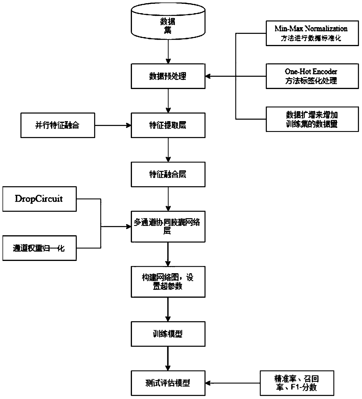
[0080] like figure 1 As shown, the main structure of the system in this implementation case includes: an image data acquisition module, an image data preprocessing module, an image feature extraction module, a network training module, and a test evaluation module; the image data acquisition module is used for crawling and collecting colon cancer. Pathological images and structured data processing; image data preprocessing module includes using Min-Max Normalization to normalize data, using One-Hot Encoder to label image categories, and using data augmentation technology to enhance data robustness properties and generalization; image feature extraction module, using weight self-adjusting feature fusion technology to extract image features; network training module, using capsule netw...


PUM


Abstract

The invention discloses a rectal cancer pathology image classification method based on a multi-channel collaborative capsule network. A dynamic routing mechanism of a capsule network is utilized to construct a network model, a multi-input feature fusion mode is utilized in a feature extraction layer to carry out feature extraction on a picture, a plurality of channels are arranged in parallel in the capsule layer to accelerate training, and then a margin loss function is utilized to train the model. The problems of spatial information loss, no rotation, translation invariance and the like in the training process of the traditional convolutional neural network technology are effectively overcome; and the generalization ability of the model is improved by utilizing feature fusion, and the capsule layer allows cooperative cooperation between channels to accelerate training so that the time cost can be effectively saved, the parameters of the network are reduced and the network training ismore efficient.

Description

technical field [0001] The invention relates to the fields of deep learning, medical image processing and computer-aided therapy, in particular to a method for classifying rectal cancer histopathological images based on a multi-channel collaborative capsule network. Background technique [0002] With the rapid development of medical technology, medical images have been greatly expanded. The scientific use of medical image analysis to efficiently and accurately classify tissue and cell images can help doctors better explore cancer treatment. Medical image analysis is one of the most basic applications and the most active research fields in recent decades. Classifying tumor tissue at the cellular level can better understand the characteristics of tumors, thereby helping their patients to better choose the means of treating cancer. Classifying tissues and cells from colon cancer images is a challenging task as cells are not limited to heterogeneity such as shape, intensity, te...

Claims


Application Information

Patent Timeline
IPC IPC(8): G06K9/46G06K9/62
CPCG06V10/40G06V2201/03G06F18/241G06F18/214
Inventor 王万良李存发屠杭垚
Owner ZHEJIANG UNIV OF TECH
Learn More