Method for detecting atrophic arc of high myopia fundus image based on machine learning

A machine learning and fundus image technology, applied in the field of medical image processing, can solve the problems of the large number of myopia patients, time-consuming and laborious, and achieve the effect of reducing the amount of calculation, improving the accuracy, and supporting the powerful auxiliary technology.

Pending Publication Date: 2020-06-05
NANJING UNIV OF AERONAUTICS & ASTRONAUTICS
View PDF2 Cites 2 Cited by
  • Summary
  • Abstract
  • Description
  • Claims
  • Application Information

AI Technical Summary

Problems solved by technology

[0003] At present, the early recognition of high myopia mainly relies on regular vision testing and the experience and judgment of doctors. However, manual testing is not only time

Method used


Image

  • Method for detecting atrophic arc of high myopia fundus image based on machine learning
  • Method for detecting atrophic arc of high myopia fundus image based on machine learning
  • Method for detecting atrophic arc of high myopia fundus image based on machine learning

Examples


Embodiment 1

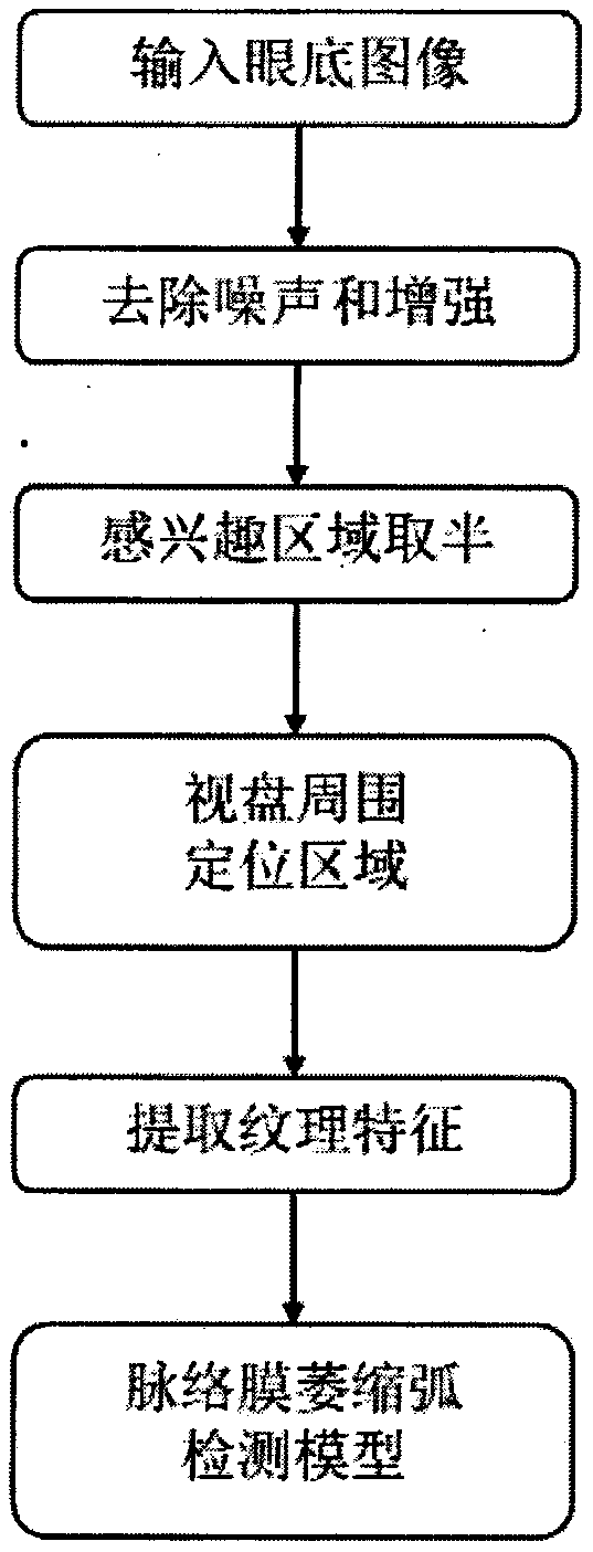
[0022] Implementation Example 1: The machine learning-based detection method of the atrophy arc of the fundus image of high myopia provided by the present invention first removes noise and enhances the input fundus image, so as to facilitate the subsequent operations of halving the region of interest and locating the optic disc region, and then Extract the features of the positioning area of ​​the optic disc and its surrounding choroidal atrophy arc, use the feature vector as the input of the choroidal atrophy arc detection model, use the gradient lifting machine in the machine learning method to classify and judge, and finally output the detection conclusion, refer to figure 1 As shown, the detection method of the atrophy arc in the fundus image of high myopia based on machine learning mainly includes the following steps:

[0023] Step 1: The local fundus image data set is used as the training and test fundus image sets, and the data set has 1200 left and right eye images of d...


PUM


Abstract

The invention discloses a method for detecting an atrophic arc of a high myopia fundus image based on machine learning, which comprises the following steps of: obtaining a fundus image, and randomly selecting a training set and a test set, removing noise and enhancing the fundus image, and extracting a main blood vessel contour, carrying out region-of-interest halving operation based on density distribution of the main blood vessels, and obtaining a positioning region of choroidal atrophic arcs of the optic disc and the periphery of the optic disc in the fundus image according to the brightness information and the shape information, taking the positioning region image as input, and extracting texture features of the positioning region image in combination with a complete local binary pattern operator and a binary Gabor pattern operator, and based on the extracted textural features, training a detection model on the training set by using a gradient elevator in machine learning, and finely adjusting parameters of the detection model to finally obtain a detection model of the atrophic arc of the high myopia fundus image. Automatic detection of choroidal atrophic arcs in fundus imagesof patients with high myopia is realized, the precision is high, and the speed is high.

Description

technical field [0001] The invention relates to a method for detecting atrophy arcs in fundus images of high myopia based on machine learning, and belongs to the field of medical image processing. Background technique [0002] The symptoms of high myopia are mainly excessive elongation of the anterior and posterior axis of the eyeball, and in the fundus, it is manifested as a choroidal atrophy arc around the optic disc, that is, a bright arc-shaped area around the optic disc that is slightly darker than the optic disc. In the fundus images of patients with high myopia, the choroid around the optic disc is pulled away from the temporal side of the nipple under the traction of the sclera, exposing the sclera behind it, forming a white arc-shaped spot. If the posterior pole of the eyeball continues to expand and extend, the detachment of the choroid gradually extends from the temporal side of the papilla to the periphery of the optic disc, and finally forms a ring-shaped spot. ...

Claims


Application Information

Patent Timeline
IPC IPC(8): G06K9/62G06K9/32G06K9/40G06K9/46
CPCG06V10/30G06V10/25G06V10/56G06V10/44G06V2201/03G06F18/2411G06F18/214
Inventor 李晗万程陈柏兵卜泽鹏叶辉
Owner NANJING UNIV OF AERONAUTICS & ASTRONAUTICS
Why Eureka
Try Eureka