
Microscopic image-based cell analysis method and device, equipment and a storage medium

A microscopic image and cell technology, applied in the field of image processing, can solve the problems of large statistical time consumption and low efficiency, and achieve the effect of shortening the statistical time

Active Publication Date: 2020-06-09
腾讯医疗健康(深圳)有限公司
View PDF10 Cites 14 Cited by
  • Summary
  • Abstract
  • Description
  • Claims
  • Application Information

AI Technical Summary

Problems solved by technology

[0004]However, the manual visual counting scheme in the related art needs to consume a lot of counting time, resulting in low efficiency of counting the number of cells and percentages of each attribute

Method used


Image

  • Microscopic image-based cell analysis method and device, equipment and a storage medium
  • Microscopic image-based cell analysis method and device, equipment and a storage medium
  • Microscopic image-based cell analysis method and device, equipment and a storage medium

Examples


Embodiment Construction

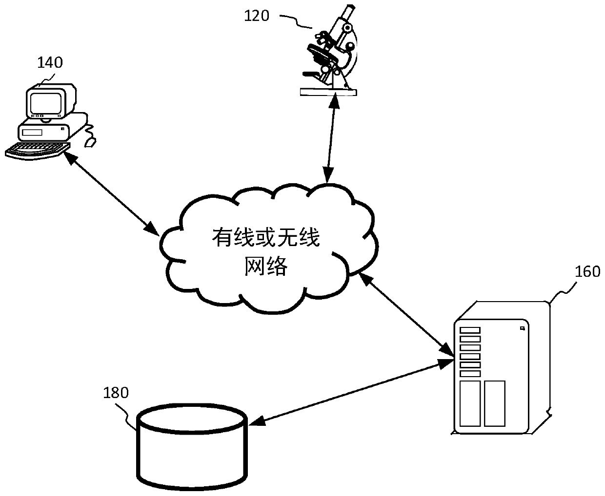
[0062] Reference will now be made in detail to the exemplary embodiments, examples of which are illustrated in the accompanying drawings. When the following description refers to the accompanying drawings, the same numerals in different drawings refer to the same or similar elements unless otherwise indicated. The implementations described in the following exemplary embodiments do not represent all implementations consistent with this application. Rather, they are merely examples of apparatuses and methods consistent with aspects of the present application as recited in the appended claims.

[0063] It should be understood that "several" mentioned herein refers to one or more, and "multiple" refers to two or more. "And / or" describes the association relationship of associated objects, indicating that there may be three types of relationships, for example, A and / or B may indicate: A exists alone, A and B exist simultaneously, and B exists independently. The character " / " gener...


PUM


Abstract

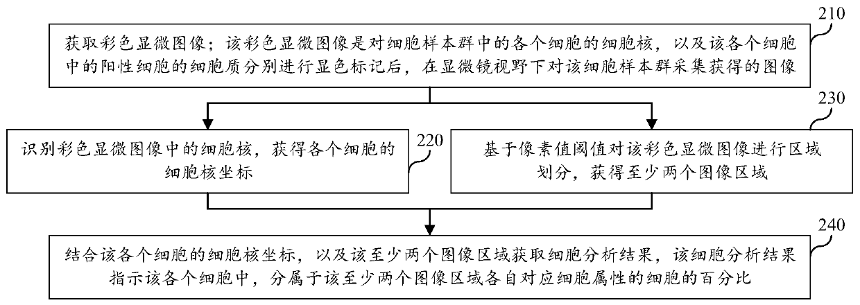
The invention relates to a microscopic image-based cell analysis method and device, equipment, and a storage medium, and relates to the technical field of image processing. The method comprises the following steps: acquiring color microscopic images for respectively displaying and marking cytoplasm of the cell nucleus and the positive cell; identifying cell nucleuses in the color microscopic imageto obtain cell nucleus coordinates of each cell; performing region division on the color microscopic image based on a pixel value threshold value; obtaining at least two image areas respectively corresponding to various cell attributes, combining cell nucleus coordinates of each cell; and at least two image regions, obtaining percentages of cells indicating various cell attributes in each cell, according to the scheme, the cell percentage is automatically counted through artificial intelligence AI, manual classified statistics is not needed, the statistical time of the cell percentage of eachattribute is greatly shortened, and the statistical efficiency of positive / negative cells in pathological diagnosis based on immunohistochemistry is improved.

Description

technical field [0001] The present application relates to the technical field of image processing, in particular to a cell analysis method, device, equipment and storage medium based on microscopic images. Background technique [0002] Immunohistochemistry is a method for assisting pathological diagnosis. It stains the cells in the cell sample population through chemical chromogenic reaction to highlight the positive cells in the cell sample population under the microscope. [0003] In pathological diagnosis based on immunohistochemistry, the number or percentage of positive cells in a cell sample population is an important basis for pathological diagnosis. In related technologies, medical staff can stain the nuclei and the cytoplasm of positive cells in the cell sample group by immunohistochemistry, and manually count the number of nuclei in the positive cells and the number of nuclei in the negative cells manually, and according to the statistical The percentage of positi...

Claims


Application Information

Patent Timeline
Patent Type & Authority Applications(China)
IPC IPC(8): G06T7/136G06T5/30G06T7/90G16H50/20
CPCG06T7/136G06T7/90G06T5/30G16H50/20G06T2207/10024G06T2207/30004
Inventor 张军田宽颜克洲姚建华韩骁
Owner 腾讯医疗健康(深圳)有限公司
Learn More