Method and system for segmenting choroidal neovascularization from fundus OCT image

A new blood vessel and choroid technology, applied in the field of medical image processing, can solve the problems of low segmentation accuracy and unclear boundary area of ​​lesions, and achieve the effect of high segmentation accuracy, clear and accurate boundary area, and accurate segmentation results.

Active Publication Date: 2020-06-16
SUZHOU UNIV
View PDF5 Cites 13 Cited by
  • Summary
  • Abstract
  • Description
  • Claims
  • Application Information

AI Technical Summary

Problems solved by technology

[0004] The purpose of the present invention is to provide a method and system for segmenting choroidal neovascularization from fundus OCT images, so as to solv...

Method used


Image

  • Method and system for segmenting choroidal neovascularization from fundus OCT image
  • Method and system for segmenting choroidal neovascularization from fundus OCT image
  • Method and system for segmenting choroidal neovascularization from fundus OCT image

Examples


Embodiment Construction

[0029] The present invention will be further described below in conjunction with the accompanying drawings. The following examples are only used to illustrate the technical solution of the present invention more clearly, but not to limit the protection scope of the present invention.

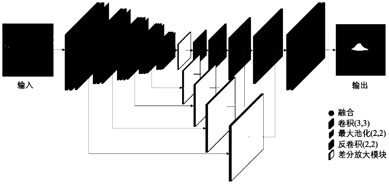
[0030] A method for segmenting choroidal neovascularization from fundus OCT images comprising,

[0031] a. Collect fundus OCT images containing choroidal neovascularization, and divide them into training set and test set;

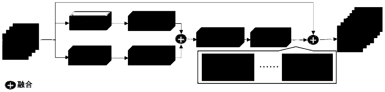
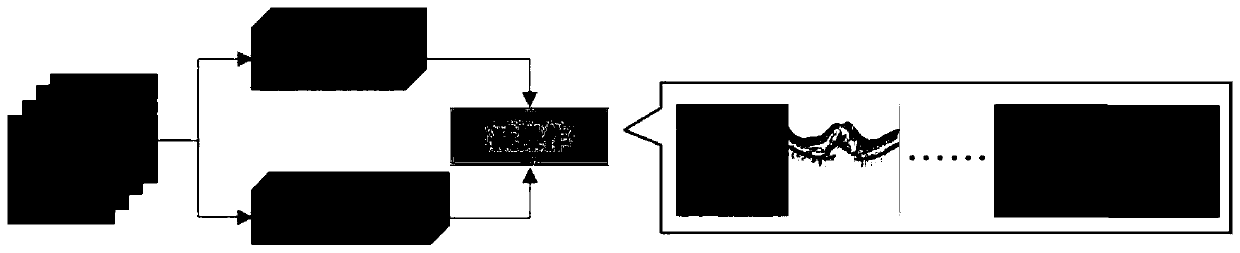
[0032] b. Construct a convolutional neural network based on the differential amplification module, using VGG16 as the feature extractor of the encoding part of the U-Net network; connect a differential amplification module after the pooling operation of each convolution block to form a skip connection, and extract High-frequency information and low-frequency information at different resolutions, the high-frequency information and the low-frequency information are respectivel...


PUM


Abstract

The invention discloses a method and system for segmenting choroidal neovascularization from a fundus OCT image in the technical field of medical image processing. The objective of the invention is tosolve the technical problems of low segmentation accuracy and unclear lesion boundary regions in a choroidal neovascularization segmentation result in the prior art. The method comprises the steps ofcollecting a fundus OCT image containing choroidal neovascularization lesions; constructing a convolutional neural network based on a differential amplification module; training and testing the constructed convolutional neural network based on the differential amplification module; and segmenting choroidal neovascularization from the fundus OCT image by using the trained network. According to theinvention, the VGG16 is used as a coding part of the U-Net network; a differential amplification module is connected after the pooling operation of each convolution block; according to the method, abinary cross entropy loss function and a Dice loss function are used as loss functions to restrain the whole network, so that the segmentation accuracy is high, and a lesion boundary region is clearerand more accurate.

Description

technical field [0001] The invention belongs to the technical field of medical image processing, and in particular relates to a method and system for segmenting choroidal neovascularization from fundus OCT images. Background technique [0002] Choroidal neovascularization (CNV) is a pathological manifestation of choroidal diseases in the retina (such as age-related macular degeneration, high myopia and other diseases), which will damage people's main vision and may lead to blindness in severe cases. Fundus fluorescein angiography (FA) and indocyanine green angiography (ICGA) are early observation methods for this disease, but since fundus images provide two-dimensional information of retinal structure, we can only observe the position and shape of CNV. The emergence of optical coherence tomography (OCT) has brought great help to the observation of the three-dimensional structure of the retina. In addition to having high resolution, it is also non-invasive imaging (it can avo...

Claims


Application Information

Patent Timeline
IPC IPC(8): G06T7/11
CPCG06T7/11G06T2207/30101G06T2207/10101
Inventor 陈新建石霏苏金珠朱伟芳
Owner SUZHOU UNIV
Why Eureka
Try Eureka