PET/CT image fusion automatic labeling method

An automatic labeling and CT image technology, applied in the field of medical mathematical image processing, can solve the problems of no obvious structural outline of the image, low image resolution, long cycle, etc., to shorten the time of attenuation correction, make up for low resolution, and accuracy high effect

Active Publication Date: 2020-06-19
ZHEJIANG SHUREN UNIV
View PDF14 Cites 1 Cited by
  • Summary
  • Abstract
  • Description
  • Claims
  • Application Information

AI Technical Summary

Problems solved by technology

[0004] However, the existing CT images require ultra-high thin-layer scanning and some examinations of auxiliary angiography in independent diagnosis to achieve a scan to determine the nature of the lesion; PET images collected in independent diagnosis have no obvious structural outlines, only in the nodules. The regional metabolism is strong, highlighted, and the image resolution is low, the cycle is long and other shortcomings;

Method used


Image

  • PET/CT image fusion automatic labeling method
  • PET/CT image fusion automatic labeling method
  • PET/CT image fusion automatic labeling method

Examples


Embodiment Construction

[0048] In order to enable those skilled in the art to better understand the technical solution of the present invention, the technical solution of the present invention will be further described below in conjunction with the accompanying drawings and embodiments.

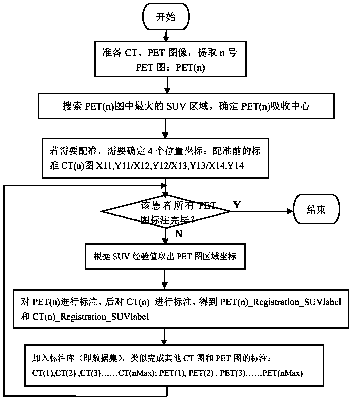
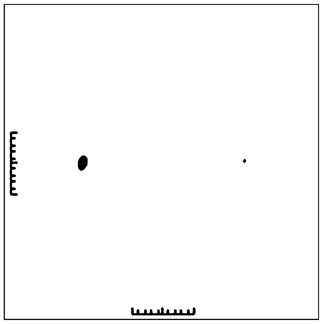
[0049] Refer to attached Picture 1-1 , Figure 1-2 , diagram 2-1 , Figure 2-2 , Figure 3-1 , Figure 3-2 , Figure 3-3 , Figure 3-4 , Pic 4-1 , Figure 4-2 , Figure 4-3 with Figure 5 As shown, a PET / CT image fusion automatic labeling method includes the following steps:

[0050] Step 1: Prepare CT and PET images. The specific steps include:

[0051] S1. Organize the sequence of CT images and PET images, and select a batch of effective images;

[0052] S2. In the selected PET image, according to the size of the SUV gray value, the serial number of the PET image with the maximum SUV gray value is automatically extracted. For example, the PET image with the serial number n is recorded as PET(n), and ...


PUM


Abstract

The invention discloses a PET/CT image fusion automatic labeling method. The method comprises the steps of: 1, preparing a CT image and a PET image; 2, automatically registering the CT image and the PET image through feature points; 3, after registration, carrying out automatic positioning on the PET image according to the SUV value, and finding out an region with violent molecular activity in thePET image; 4, carrying out region growth on a region with violent molecular activity in the PET image through a region growth method so as to realize labeling of a molecular active region; 5, fusingthe CT image and the PET image according to requirements, labeling the outline in the PET image into the CT image, and finally generating a PET/PC fusion image and a labeled CT image. After the CT image and the PET image are fused, the defects of the PET image and the CT image when the PET image and the CT image are used independently are overcome; meanwhile, the lesion focus structure and the functional metabolism condition of lesion are shown on one image at the same time, and a doctor can diagnose diseases and determine a treatment scheme conveniently.

Description

technical field [0001] The invention relates to the technical field of medical mathematical image processing, in particular to an automatic labeling method for PET / CT image fusion. Background technique [0002] CT scans a layer of a certain thickness of a certain part of the human body with an X-ray beam. The X-rays that pass through this layer are received by the detector and converted into visible light, which is converted from photoelectricity into an electrical signal, and then passed through an analog / digital converter ( Digital / Analog Converter) into numbers, convert each number in the digital matrix into small squares with grayscales ranging from black to white, that is, pixels (Pixel), and arrange them in a matrix to form a CT image, including data acquisition , the process from X-ray generation to information data acquisition; data processing, the A / D (Digital / Analog Converter) converter converts the analog signal into a digital signal and becomes the original image...

Claims


Application Information

Patent Timeline
Patent Type & Authority Applications(China)
IPC IPC(8): G16H30/40G06T5/50G06T7/33G06T7/66A61B6/00A61B6/03A61B6/12
CPCG16H30/40G06T5/50G06T7/33G06T7/66A61B6/037A61B6/032A61B6/12A61B6/5235G06T2207/10081G06T2207/10104G06T2207/20221
Inventor 白直灿叶时平陈华锋陈超祥许娅芬戴国勇
Owner ZHEJIANG SHUREN UNIV
Why Eureka
Try Eureka