Mammary gland X-ray image analysis method and device based on joint symptoms

An X-ray and symptom technology, applied in the field of image analysis, can solve problems such as low accuracy rate and weak interpretation, and achieve the effect of improving accuracy

Pending Publication Date: 2020-06-23
BEIJING SHENRUI BOLIAN TECH CO LTD +1
View PDF8 Cites 3 Cited by
  • Summary
  • Abstract
  • Description
  • Claims
  • Application Information

AI Technical Summary

Problems solved by technology

[0003] In view of this, the embodiment of the present application provides a combined sign-based mammogram image analysis method and device, which solves the problems of poor interpretability and low accuracy of existing mammogram image analysis methods

Method used


Image

  • Mammary gland X-ray image analysis method and device based on joint symptoms
  • Mammary gland X-ray image analysis method and device based on joint symptoms
  • Mammary gland X-ray image analysis method and device based on joint symptoms

Examples


Embodiment Construction

[0026] The following will clearly and completely describe the technical solutions in the embodiments of the application with reference to the drawings in the embodiments of the application. Apparently, the described embodiments are only some, not all, embodiments of the application. Based on the embodiments in this application, all other embodiments obtained by persons of ordinary skill in the art without creative efforts fall within the protection scope of this application.

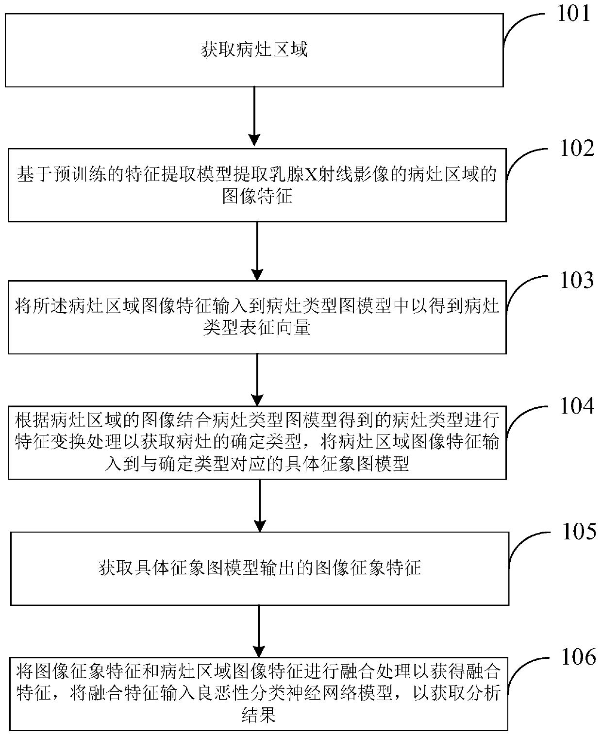
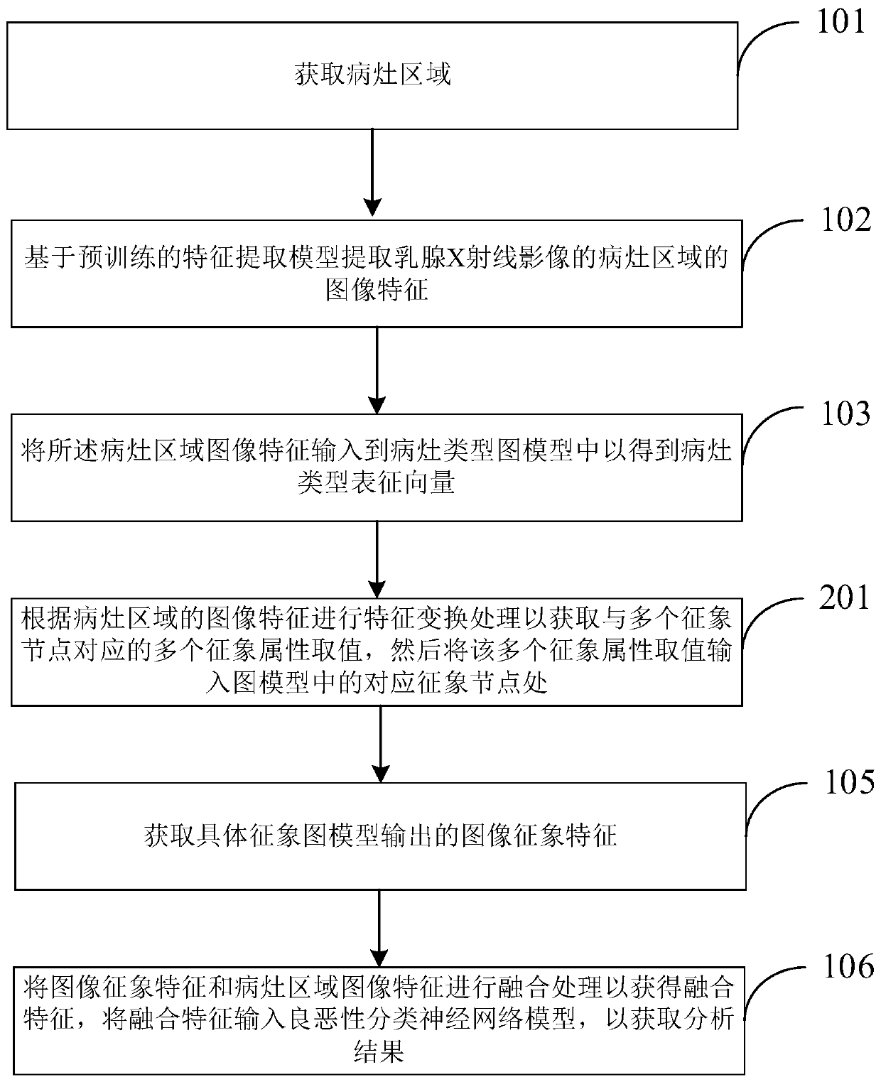
[0027] figure 1 Shown is a schematic flowchart of a method for analyzing mammograms based on combined signs provided by an embodiment of the present application. Such as figure 1 As shown, the mammography X-ray image analysis method includes the following steps:

[0028] Step 101: Obtain the lesion area. The lesion area can be obtained based on annotations, or by using any general detection and segmentation algorithm.

[0029] Step 102: Extract image features of the lesion area of ​​the mammogram bas...


PUM


Abstract

The embodiment of the invention provides a mammary gland X-ray image analysis method and device based on joint symptoms. The method comprises the steps that a focus area is acquired; extracting focusarea image features of the mammary gland X-ray image based on a pre-trained feature extraction model; inputting the lesion area image features into a lesion type graph model to obtain a lesion type representation vector; feature transformation processing is performed according to the focus area image features in combination with the focus type obtained by the focus type graph model so as to obtainthe determined type of the focus; inputting the focus area image features into a specific symptom graph model corresponding to the determined type, the specific symptom graph model comprising a plurality of symptom nodes, and at least two symptom nodes in the plurality of symptom nodes being connected through a connecting line used for representing relevancy; and obtaining image symptom featuresoutput by the specific symptom graph model.

Description

technical field [0001] The present application relates to the technical field of image analysis, in particular to a method, device, electronic equipment and computer-readable storage medium for mammography image analysis based on combined signs. Background technique [0002] Breast cancer is currently the most common cancer among women worldwide. Mammography is the most important means of early screening of breast cancer. When X-rays pass through the human body, they are absorbed in different degrees, and the amount of X-rays after passing through the human body is different. In this way, the formed image carries the information of the density distribution of various parts of the human body, and the fluorescent effect caused on the fluorescent screen or photographic film Or there is a big difference in the strength of the photosensitive effect, so shadows of different densities will be shown on the fluorescent screen or on the photographic film (after developing and fixing)...

Claims


Application Information

Patent Timeline
Patent Type & Authority Applications(China)
IPC IPC(8): G06T7/00
CPCG06T7/0012G06T2207/10116G06T2207/30068G06T2207/20081G06T2207/20084G06T2207/30096
Inventor 王楚然刚亚栋张番栋张笑春俞益洲王亦洲
Owner BEIJING SHENRUI BOLIAN TECH CO LTD
Why Eureka
Try Eureka