Intraoperative positioning method, device, storage medium and electronic device for intraoperative preset area

A technology of preset area and positioning method, which is applied in the field of image processing, can solve the problems of high dosage, limited surgical environment, and inability to meet the needs of accurate, convenient and efficient positioning, and achieve the effect of efficient positioning

Active Publication Date: 2021-06-29
PERCEPTION VISION MEDICAL TECH CO LTD +1
View PDF6 Cites 0 Cited by
  • Summary
  • Abstract
  • Description
  • Claims
  • Application Information

AI Technical Summary

Problems solved by technology

[0004] Based on this, the inventors of the present application found that although intraoperative CT positioning is accurate, safe, and fast, the dose used is high, and surgical scenarios that require multiple positioning will lead to accumulation of patient exposure doses; equipment usage scenarios are limited , cannot be installed in unshielded spaces such as operating rooms, and has great restrictions on shuttles in CT shielded rooms and sterile surgical environments; the equipment is expensive and cannot be popularized in conventional surgical departments
Unable to meet the precise, convenient and efficient positioning requirements in surgery

Method used


Image

  • Intraoperative positioning method, device, storage medium and electronic device for intraoperative preset area
  • Intraoperative positioning method, device, storage medium and electronic device for intraoperative preset area
  • Intraoperative positioning method, device, storage medium and electronic device for intraoperative preset area

Examples


Embodiment Construction

[0021] The specific embodiments of the present invention will be described in detail below with reference to the accompanying drawings, but it should be understood that the protection scope of the present invention is not limited by the specific embodiments.

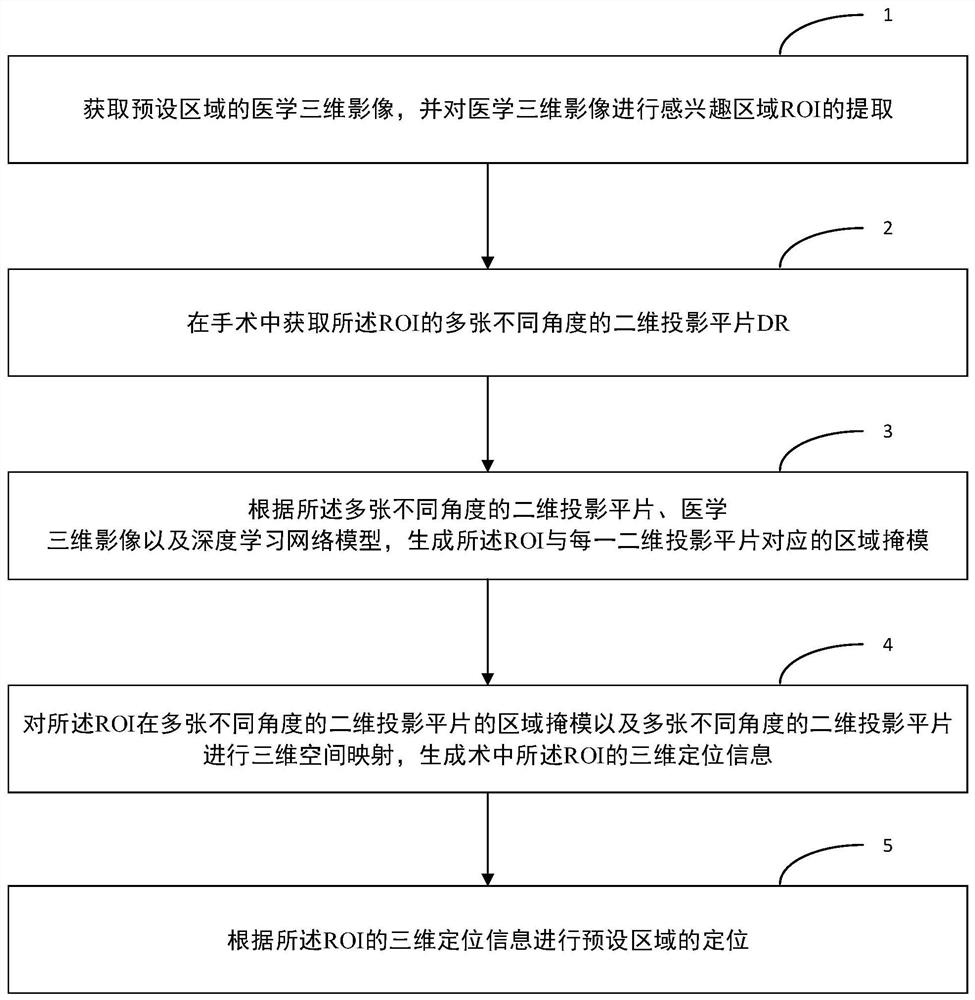
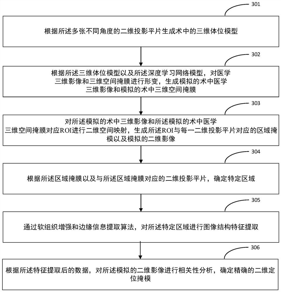
[0022] Digital radiography (DR) is currently the most widely used X-ray imaging equipment. For surgical positioning scenarios, portable DR such as mobile DR or C-arm is generally used in orthopaedics or interventional therapy. It has high spatial resolution and imaging speed. However, it can only provide two-dimensional tissue stacking images, and often cannot directly observe small lesions with small differences in tissue density. On the other hand, due to breathing motion, positioning error and posture differences, there are often large differences in the posture of intraoperative DR photography and preoperative diagnostic imaging, and the spatial mapping relationship from medical 3D images to 2D projections cannot be d...


PUM


Abstract

The invention discloses a positioning method, device, storage medium and electronic equipment for a preset area in an operation. The method includes: acquiring a medical three-dimensional image of the preset area, and extracting an ROI of a region of interest from the medical three-dimensional image; Obtain multiple 2D projection plain films from different angles of the ROI; generate a region mask corresponding to the ROI and each 2D projection plain film based on multiple 2D projection plain films from different angles, medical 3D images, and deep learning network models ; Perform 3D space mapping on the area mask of the ROI in multiple 2D projection plain films at different angles and multiple 2D projection plain films at different angles to generate 3D positioning information of the ROI in the operation for the positioning of the preset area . The positioning method, device, storage medium and electronic equipment of the intraoperative preset area provided by the present invention can realize accurate three-dimensional positioning, solve the problem of excessively high dosage in conventional surgical positioning in the prior art, and realize accurate and convenient operation , Efficient positioning requirements.

Description

technical field [0001] The present invention relates to the technical field of image processing, and in particular, to a method, device, storage medium and electronic device for locating an intraoperative preset area. Background technique [0002] Refinement operations such as puncture, interventional and minimally invasive procedures are currently widely used in clinical examination and treatment. For internal organ lesions (such as nodules, tumors and other regions of interest) that have no clear boundary and cannot be directly observed, the current main method is to use computer X-ray tomography (full name: computed tomography, English abbreviation: CT) or magnetic The three-dimensional image of the resonance image (English full name: Magnetic Resonance, English abbreviation: MR) is used for preoperative diagnosis. In the process of refined surgery such as puncture, intervention and minimally invasive after diagnosis, precise positioning and precise treatment are require...

Claims


Application Information

Patent Timeline
Patent Type & Authority Patents(China)
IPC IPC(8): G06T17/00G06T7/73G06T7/40G06T7/136G06T7/13G06T7/11G06T7/00G06T3/40G06T3/00G06K9/32
CPCG06T17/00G06T3/005G06T3/4007G06T7/11G06T7/136G06T7/13G06T7/73G06T7/40G06T7/0012G06T2207/10081G06T2207/30008G06V10/25
Inventor 魏军谢培梁赖耀明罗召洋吕丽云
Owner PERCEPTION VISION MEDICAL TECH CO LTD
Why Eureka
Try Eureka