
UNET-based cervical pathological tissue segmentation method

A cervical and pathological technology, applied in the field of cervical pathological tissue segmentation based on UNET, can solve the problems of being unable to be used in clinical practice and long processing time, and achieve the effects of reducing workload, reducing detection costs, and huge economic and social benefits

Pending Publication Date: 2020-07-31
WUHAN LANDING INTELLIGENCE MEDICAL CO LTD
View PDF1 Cites 7 Cited by
  • Summary
  • Abstract
  • Description
  • Claims
  • Application Information

AI Technical Summary

Problems solved by technology

[0004] For very large images, the above method has not been used in clinical practice due to the long processing time

Method used


Image

  • UNET-based cervical pathological tissue segmentation method
  • UNET-based cervical pathological tissue segmentation method
  • UNET-based cervical pathological tissue segmentation method

Examples


Embodiment Construction

[0048] The following will clearly and completely describe the technical solutions in the embodiments of the present invention with reference to the accompanying drawings in the embodiments of the present invention. Obviously, the described embodiments are only some, not all, embodiments of the present invention. Based on the embodiments of the present invention, all other embodiments obtained by persons of ordinary skill in the art without making creative efforts belong to the protection scope of the present invention.

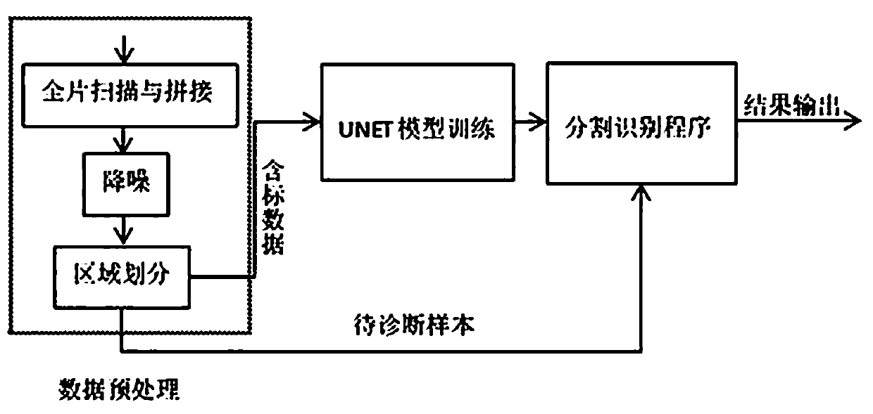
[0049] Please refer to Figures (1-3), the present invention provides the following technical solutions: a novel overlapping segmentation method for exfoliated epithelial cells, including five steps of scan stitching, noise reduction, region division, UNET model training and auxiliary diagnosis, the steps are as follows :

[0050] Step 1. Scanning stitching: first adjust the optical microscope to the 10x objective lens to scan all the marked sample smears, and ...


PUM


Abstract

The invention discloses a UNET-based cervical pathological tissue segmentation method. The method comprises five steps of scanning splicing, noise reduction, region division, UNET model training and auxiliary diagnosis. According to the method, the UNET (a convolutional neural network model adopting a deep automatic coding and decoding structure) is applied to effectively solve the common problemsthat in the field of cervical tissue pathology digital image intelligent analysis, the number of samples is small, the standardization of the samples is poor, and the real-time performance of sampleprocessing is poor, so that clinical application is hindered. The method can also help doctors to quickly and accurately identify diseased tissues and give auxiliary diagnosis suggestions, so that notonly is the workload of pathologists greatly reduced, but also the detection cost is remarkably reduced, and the method has huge economic benefits and social benefits.

Description

technical field [0001] The invention relates to the technical field of medical image segmentation, in particular to a UNET-based cervical pathological tissue segmentation method. Background technique [0002] Cervical tissue biopsy is the last link in the diagnosis of cervical diseases. In the past, pathologists have been giving the final diagnosis after manual observation. Due to the influence of expert technical experience and subjectivity, the accuracy of the diagnosis results needs to be improved. With the maturity of digital microscopic imaging technology and the new progress of artificial intelligence, it is the general trend to use computers to analyze sample slices, assist doctors in making a diagnosis, and reduce the workload of doctors. [0003] At present, the fully automatic analysis of digital images of cervical tissue pathology in the world is mainly realized through traditional machine learning. , and then use graph theory to find the central axis, rotate and...

Claims


Application Information

Patent Timeline
IPC IPC(8): G06T7/11G06T7/13G06T7/187G06T7/194G06T7/62G06T5/00G16H50/20
CPCG06T7/11G06T7/13G06T7/187G06T7/194G06T7/62G16H50/20G06T2207/10061G06T2207/20081G06T2207/30096G06T5/70Y02A90/10
Inventor 谢艾纾段慧芳庞宝川刘娟孙小蓉
Owner WUHAN LANDING INTELLIGENCE MEDICAL CO LTD
Learn More