Joint synovial membrane biopsy needle structure and usage method thereof

A biopsy needle and synovial membrane technology, applied in the field of medical devices, can solve the problems of difficulty in diagnosing arthritis types, difficult to remove synovial tissue, and high risk of infection, and achieve the effects of ingenious structure, improved adhesion rate, and improved accuracy rate.

Pending Publication Date: 2020-08-07
杜望磊 +1
View PDF9 Cites 0 Cited by
  • Summary
  • Abstract
  • Description
  • Claims
  • Application Information

AI Technical Summary

Problems solved by technology

[0003] The traditional synovial biopsy operation is traumatic and has a high risk of infection. The puncture of the joint cavity can only extract the joint fluid, and it is difficult to remove the syno

Method used


Image

  • Joint synovial membrane biopsy needle structure and usage method thereof
  • Joint synovial membrane biopsy needle structure and usage method thereof
  • Joint synovial membrane biopsy needle structure and usage method thereof

Examples


Embodiment Construction

[0018] In the following, numerous specific details are set forth in order to provide a thorough understanding of the concepts underlying the described embodiments. It will be apparent, however, to one skilled in the art that the described embodiments may be practiced without some or all of these specific details. In other instances, well known processing steps have not been described in detail.

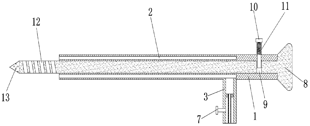
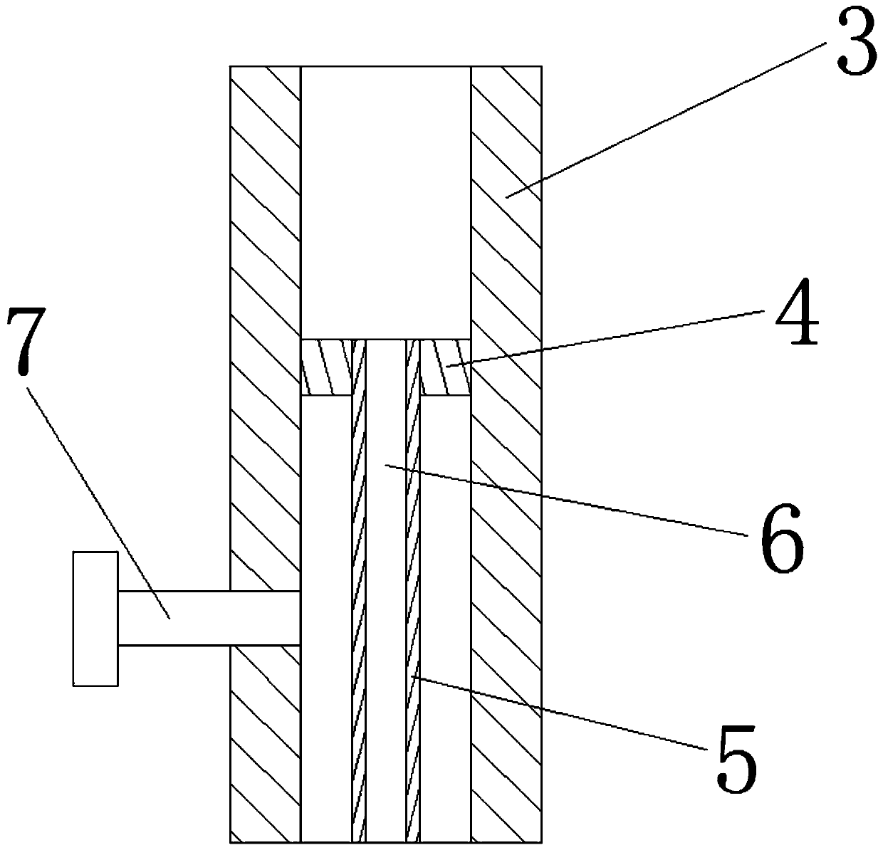
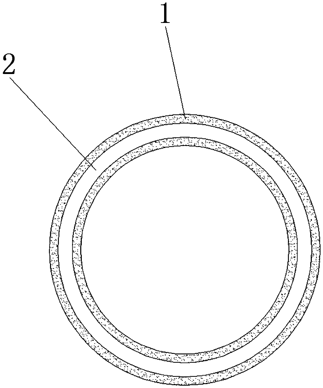
[0019] Such as figure 1 , figure 2 , image 3 As shown, the structure of the joint synovial biopsy needle includes a sleeve 1, a cavity 2, a connecting sleeve 3, a mounting plate 4, a connecting pin 5, a drain hole 6, a bolt 7, a puncture needle 8, a positioning hole 9, and a sliding pin 10. Spring 11, thread blade 12, tapered needle 13, the cavity 2 surrounds the inside of the sleeve 1, the cavity 2 is integrally connected with the sleeve 1, and the connecting sleeve 3 runs through the sleeve 1 On the right side of the bottom, the connecting sleeve 3 is connected with the sleeve...


PUM


Abstract

The invention discloses a joint synovial membrane biopsy needle structure and a usage method thereof. The joint synovial membrane biopsy needle structure comprises a sleeve, a cavity, a connecting sleeve, a mounting plate, a connecting pin, a liquid discharging hole, a bolt, a puncture needle, a positioning hole, a sliding pin, a spring, a threaded blade and a conical needle head, the invention discloses a joint synovial membrane biopsy needle structure and a usage method thereof. The synovial fluid extractor is ingenious in structure, powerful in function and easy to operate, joint effusion,namely synovial fluid, can be extracted by using the synovial fluid extractor, synovial membranes can also be extracted, the synovial fluid extractor has an instructive effect on diagnosis and treatment of arthritis, and in addition, synovial membrane tissue extraction plays a huge role in promoting scientific research of arthritis in scientific research.

Description

technical field [0001] The invention relates to the technical field of medical devices, in particular to a structure of a joint synovial biopsy needle and a method for using the same. Background technique [0002] The synovial membrane (synovial cell) is the inner layer of the joint capsule, light red, smooth and shiny, thin and soft, composed of loose connective tissue. The connective tissue membrane that covers the inside of the joint capsule. [0003] The traditional synovial biopsy operation is traumatic and has a high risk of infection. The puncture of the joint cavity can only extract the joint fluid, and it is difficult to remove the synovial tissue, which makes it difficult to diagnose the type of arthritis. In view of the above defects, it is necessary to design a synovial biopsy. Structure of a membrane biopsy needle and method of use thereof. Contents of the invention [0004] The technical problem to be solved by the present invention is to provide a structur...

Claims


Application Information

Patent Timeline
IPC IPC(8): A61B10/00A61B10/02
CPCA61B10/0045A61B10/0266A61B10/0283
Inventor 杜望磊杜米娜杨西超赵金康吴振彪
Owner 杜望磊
Why Eureka
Try Eureka