
Detection device used for fish kidney tissue cells

A technology of tissue cells and detection devices, which is applied in the direction of measuring devices, biochemical cleaning devices, enzymology/microbiology devices, etc., can solve the problems of microscope body blocking and inconvenient, and achieve the effect of convenient disassembly, observation and detection

Pending Publication Date: 2020-08-14
CHONGQING INST OF GREEN & INTELLIGENT TECH CHINESE ACADEMY OF SCI +1
View PDF12 Cites 2 Cited by
  • Summary
  • Abstract
  • Description
  • Claims
  • Application Information

AI Technical Summary

Problems solved by technology

The cell assay detection device for kidney transplantation in the above-mentioned patents has the following disadvantages: when disassembling or fixing cell slices during use, there is a problem that the microscope body is blocked and inconvenient

Method used


Image

  • Detection device used for fish kidney tissue cells
  • Detection device used for fish kidney tissue cells
  • Detection device used for fish kidney tissue cells

Examples


Embodiment 1

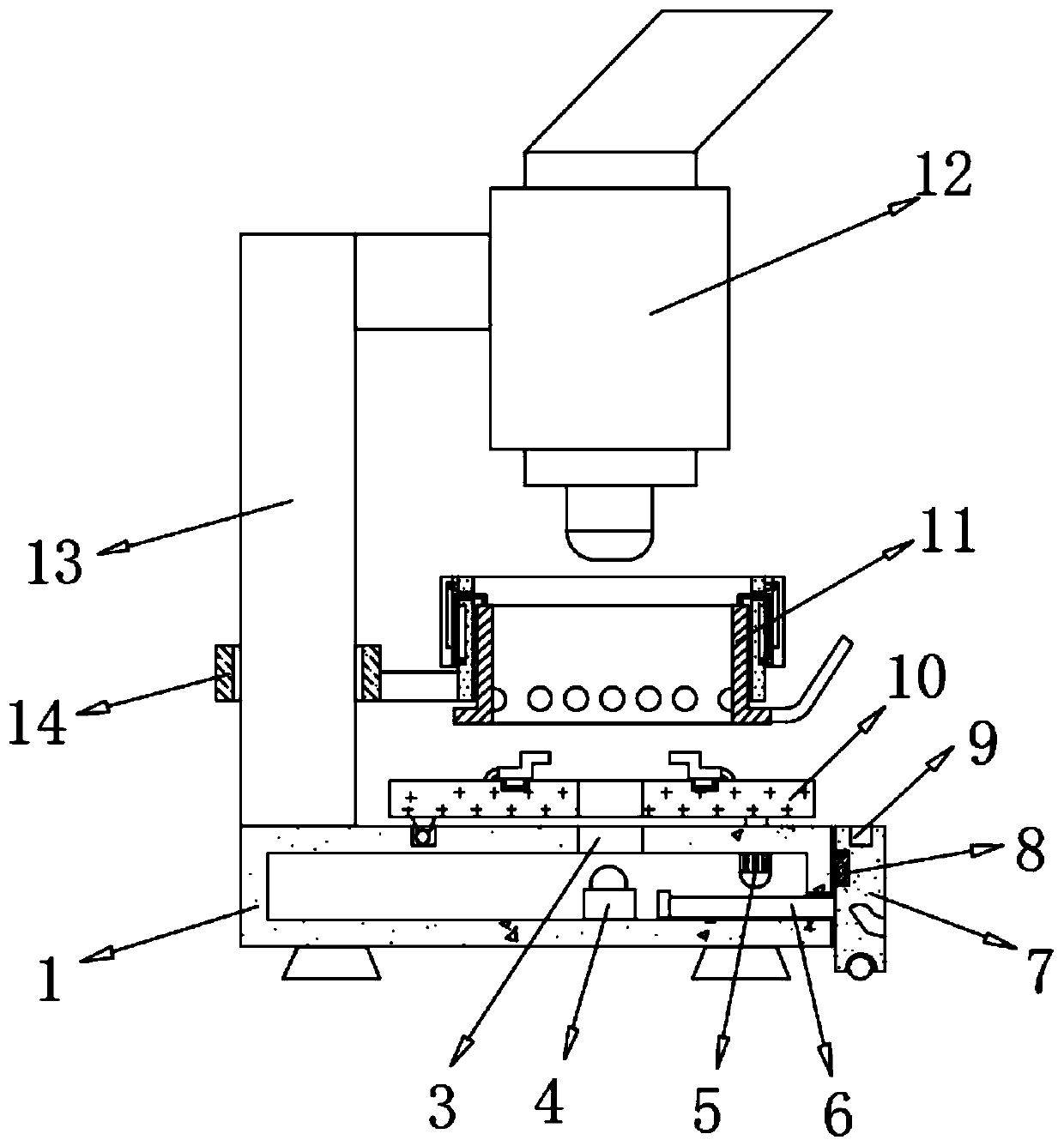
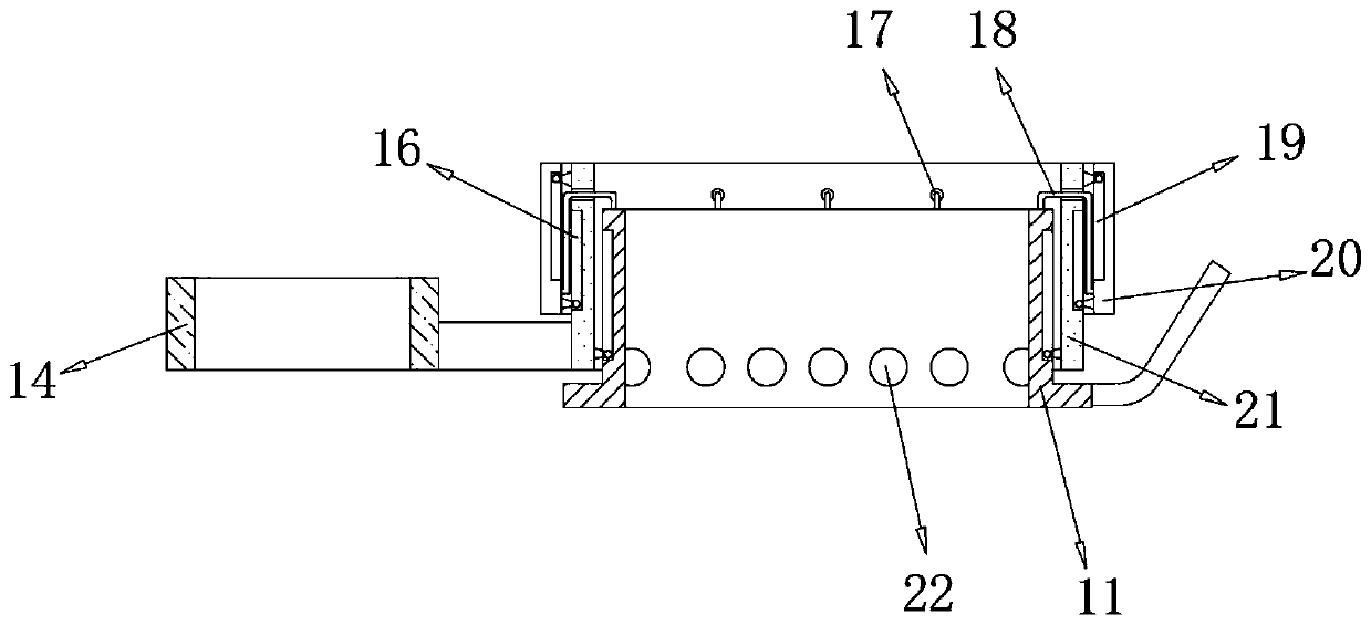
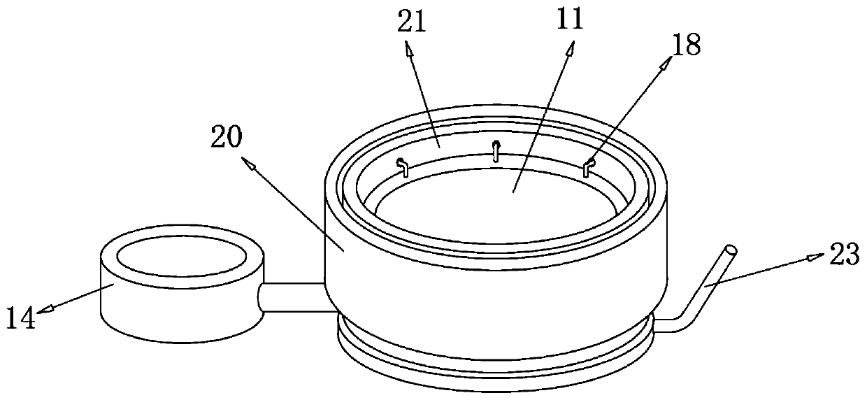
[0029] refer to Figure 1-3 , a detection device for fish kidney tissue cells, comprising a base 1, the left side of the top outer wall of the base 1 is connected with a column 13 by fastening bolts, and the top of the column 13 is connected with a microscope body 12 by fastening bolts, the base 1. The right side of the inner wall of the top is connected with the rotary motor 5 through fastening bolts, and the output shaft of the rotary motor 5 is connected with the placement plate 10 through the fastening bolts. A perforation 3, and the bottom inner wall of the base 1 is connected with the first illuminating lamp 4 through fastening bolts, and the placing plate 10 can be driven by the rotating motor 5 to rotate out of the bottom of the microscope body 12, and the fish kidney slice cells are placed on the placing plate 10 At the 3 first perforations, after the fixation is completed, the rotating motor 5 drives the placement plate 10 to rotate to the bottom of the microscope bo...

Embodiment 2

[0040] refer to Figure 4-5 , a warp knitting machine pulling and winding device for weaving plastic nets. Compared with Embodiment 1, this embodiment has a groove 2 on one side of the placement board 10, and the column 13 is close to the side of the placement board 10. The bottom is connected with a limiting rod 15 by fastening bolts, and the outer diameter of the limiting rod 15 is compatible with the inner diameter of the groove 2 .

[0041] Working principle: when the rotating motor 5 drives the placing plate 10 to rotate and reset, the limit rod 15 moves in the groove 2 to limit the reset of the placing plate 10 .


PUM


Abstract

The invention discloses a detection device used for fish kidney tissue cells. The detection device comprises a base, wherein a stand column is arranged on the left side of the outer wall of the top ofthe base; a microscope body is arranged on the top of the stand column; a rotating motor is arranged on the right side of the inner wall of the top of the base, and a placement plate is arranged on an output shaft of the rotating motor; first penetrating holes are formed in the circle center of the top of the placement plate and the outer wall of the top of the base; and a first illuminating lampis arranged on the inner wall of the bottom of the base. The placement plate can be driven by the rotating motor to rotate out of the bottom of the microscope body, fish kidney slice cells are placedat the first penetrating holes in the placement plate, the placement plate is driven by the rotating motor to rotate to the bottom of the microscope body for observation after fixation is completed,and meanwhile the first penetrating holes are illuminated by the first illuminating lamp, so that observation and detection are facilitated, cell tissues are convenient to disassemble and assemble, the specific position of the cell section does not need to be adjusted every time, and convenience and high efficiency are achieved.

Description

technical field [0001] The invention relates to the technical field of fish cell detection, in particular to a detection device for fish kidney tissue cells. Background technique [0002] In the detection of fish kidney tissue cells, a microscope is needed for observation and detection. The existing microscopes have the problem of inconvenient fixation and disassembly of cell slices. [0003] After searching, the Chinese patent publication number is CN205443295U, which discloses a cell assay detection device for kidney transplantation, including a device body, which includes a base, a column, a beam, a tight seat, and a lens barrel, and the bottom end of the column is fixed. The base is connected, the upper end of the column is movably connected to a plurality of crossbeams, each crossbeam is fixedly connected to a holding seat, the holding seat is fixedly connected to the lens barrel, the upper end of the lens barrel is fixedly connected to the eyepiece, and the lower end o...

Claims


Application Information

Patent Timeline
Patent Type & Authority Applications(China)
IPC IPC(8): C12M1/34C12M1/00
CPCG01N33/483
Inventor 李天云裴得胜贾盼盼李勇志
Owner CHONGQING INST OF GREEN & INTELLIGENT TECH CHINESE ACADEMY OF SCI
Learn More