Adenocarcinoma pathological image analysis method based on priori perception and multi-task learning

A multi-task learning, pathological image technology, applied in the field of adenocarcinoma pathological image analysis based on prior perception and multi-task learning, can solve problems such as lack of pathological level interpretation, and achieve high automatic diagnosis accuracy and robustness. The effect of strengthening and reducing physical strength

Pending Publication Date: 2020-08-18
NANJING UNIV OF INFORMATION SCI & TECH
View PDF0 Cites 1 Cited by
  • Summary
  • Abstract
  • Description
  • Claims
  • Application Information

AI Technical Summary

Problems solved by technology

Although it reaches or even exceeds the performance of human level, it lacks the explanation of pathological level when it is applied clinically

Method used


Image

  • Adenocarcinoma pathological image analysis method based on priori perception and multi-task learning
  • Adenocarcinoma pathological image analysis method based on priori perception and multi-task learning
  • Adenocarcinoma pathological image analysis method based on priori perception and multi-task learning

Examples


specific Embodiment 1

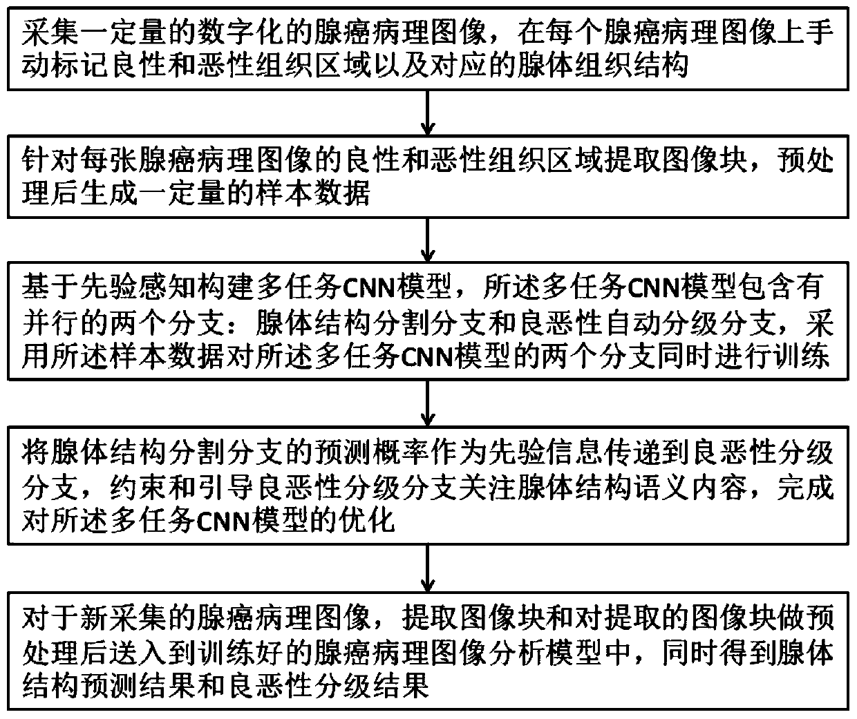
[0044] combine figure 1 , the present invention proposes an adenocarcinoma pathological image analysis method based on prior perception and multi-task learning, the image analysis method comprising:

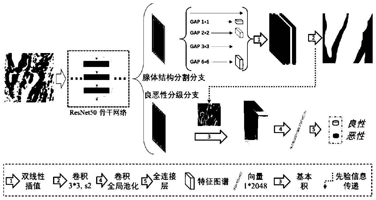
[0045]For the newly collected adenocarcinoma pathological image, the image block is extracted and the extracted image block is preprocessed and sent to the trained adenocarcinoma pathological image analysis model based on the multi-task learning method, the adenocarcinoma pathological image analysis model The model contains two parallel branches: glandular structure segmentation branch and benign and malignant automatic grading branch, and the prediction results of glandular structure and benign and malignant grading results are obtained at the same time.

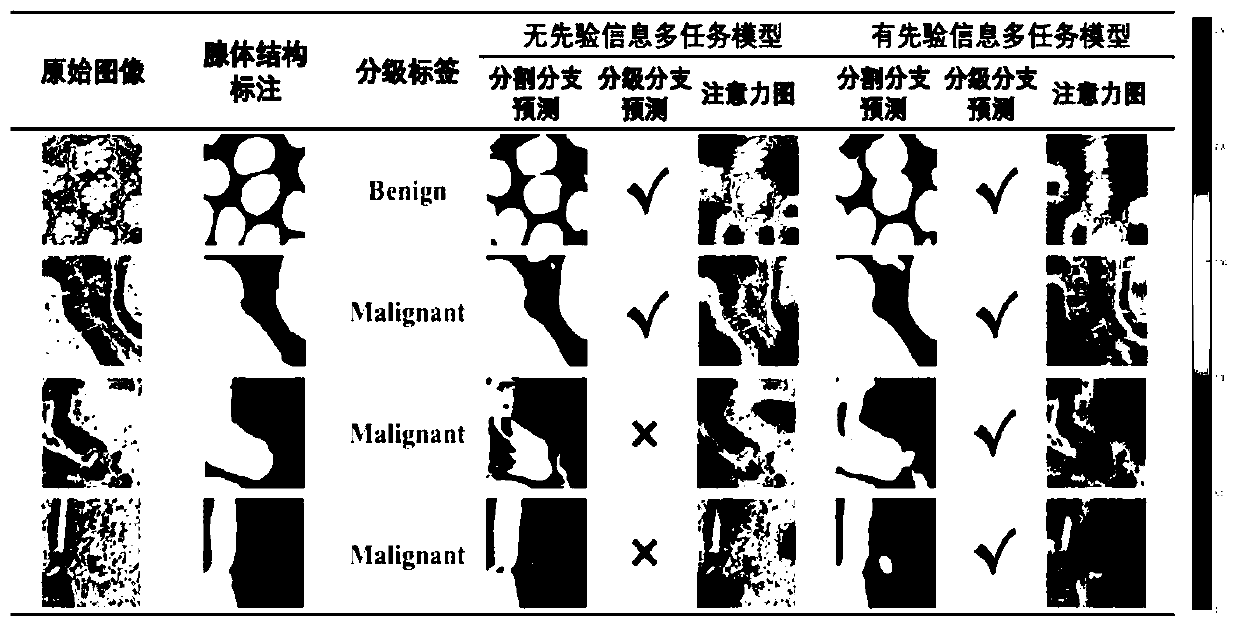
[0046] Wherein, the segmentation result of the gland structure segmentation branch is transmitted to the benign and malignant automatic grading branch as prior information, and the benign and malignant grading branch is constr...

specific Embodiment 2

[0061] The present invention also mentions an adenocarcinoma pathological image analysis device based on prior perception and multi-task learning, the image analysis device includes an adenocarcinoma pathological image preprocessing module, an adenocarcinoma pathological image analysis model, and an adenocarcinoma pathological image analysis model building blocks.

[0062] The adenocarcinoma pathological image preprocessing module is used for performing corresponding preprocessing on newly acquired adenocarcinoma pathological images, and the preprocessing includes extracting image blocks and image block enhancement processing. Preferably, the adenocarcinoma pathological image preprocessing module can preprocess the sample image and the newly acquired image at the same time, reducing redundant functional modules.

[0063] The adenocarcinoma pathological image analysis model includes two parallel branches: a glandular structure segmentation branch and a benign and malignant auto...

specific Embodiment 3

[0069] The present invention also mentions an adenocarcinoma pathological image analysis system based on prior perception and multi-task learning, and the image analysis system includes a processor and a memory connected to each other.

[0070] A computer execution program is stored in the memory, and the processor executes the computer execution program stored in the memory to execute the adenocarcinoma pathological image analysis method based on prior perception and multi-task learning as described above.

[0071] Aspects of the invention are described in this disclosure with reference to the accompanying drawings, which show a number of illustrated embodiments. Embodiments of the present disclosure are not necessarily defined to include all aspects of the present invention. It should be appreciated that the various concepts and embodiments described above, as well as those described in more detail below, can be implemented in any of numerous ways, since the concepts and emb...


PUM


Abstract

The invention discloses an adenocarcinoma pathological image analysis method based on priori perception and multi-task learning. The method comprises the following steps: for a newly acquired adenocarcinoma pathological image, extracting image blocks, preprocessing the extracted image blocks, and sending the preprocessed image blocks into a trained adenocarcinoma pathological image analysis modelconstructed based on a multi-task learning method, wherein the adenocarcinoma pathological image analysis model comprises two parallel branches: a gland structure segmentation branch and a benign andmalignant automatic grading branch, and obtaining a gland structure prediction result and a benign and malignant grading result at the same time. The segmentation result of the gland structure segmentation branch is used as prior information to be transmitted to the benign and malignant automatic grading branch, and the benign and malignant grading branch is restrained and guided to grade the adenocarcinoma pathological image after paying attention to the semantic content of the gland structure. According to the method, parallel gland tissue segmentation and automatic grading can be realized,the physical strength, energy and time cost investment of manual diagnosis of pathologists is reduced, and the accuracy of automatic diagnosis is improved.

Description

technical field [0001] The present invention relates to the deep learning method of artificial intelligence and the technical field of medical pathology, in particular to an adenocarcinoma pathological image analysis method, device and system based on prior perception and multi-task learning. Background technique [0002] With the development of computer vision technology, more and more advanced image processing algorithms have been applied to the field of medical images. In the field of digital pathology, deep learning is playing an increasingly important role due to its excellent performance in image classification, tissue segmentation, and cell detection. Usually, deep learning methods simply implement the processing of image pixels for final label prediction. Although it reaches or even exceeds the performance of human level, it lacks the explanation of pathological level when it is applied clinically. Therefore, this needs to consider the prior knowledge of pathology ...

Claims


Application Information

Patent Timeline
Patent Type & Authority Applications(China)
IPC IPC(8): G16H30/40G16H70/60G06K9/62G06N3/08G06T7/11
CPCG16H30/40G16H70/60G06T7/11G06N3/08G06F18/214
Inventor 闫朝阳徐军
Owner NANJING UNIV OF INFORMATION SCI & TECH
Why Eureka
Try Eureka