Intravascular image fusion method, intravascular image fusion system and image acquisition device

An image acquisition device and image fusion algorithm technology, applied in the field of medical imaging, can solve the problems of low quality of image fusion results and incomplete details, and achieve high-quality results

Inactive Publication Date: 2020-09-15
QUANJING HENGSHENG BEIJING SCI & TECH CO LTD
View PDF10 Cites 12 Cited by
  • Summary
  • Abstract
  • Description
  • Claims
  • Application Information

AI Technical Summary

Problems solved by technology

Although this patent proposes to fuse IVUS images and OCT images, it only fuses IVUS images and OCT images according to the outline, and there are also disadvantages such as low quality of image fusion results and incomplete details.

Method used


Image

  • Intravascular image fusion method, intravascular image fusion system and image acquisition device
  • Intravascular image fusion method, intravascular image fusion system and image acquisition device
  • Intravascular image fusion method, intravascular image fusion system and image acquisition device

Examples


Embodiment Construction

[0055] The technical solution of the present invention will be further described in detail below in conjunction with specific examples, but the protection scope of the present invention is not limited to the following description.

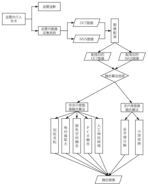
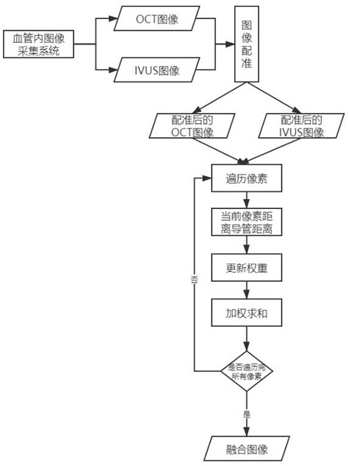
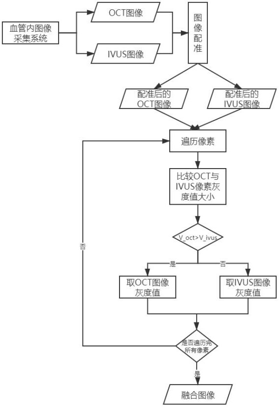
[0056] Such as figure 1 Shown, a kind of intravascular image fusion method, this method comprises:

[0057] Simultaneously collecting a first modality image and a second modality image in the blood vessel, wherein the first modality image and the second modality image belong to different types of modality images;

[0058] An image fusion algorithm is used to fuse the first mode image and the second mode image to obtain a corresponding fusion image.

[0059] In one aspect, the present invention collects images of two different modalities at the same time in the same part of the blood vessel to obtain the first modal image and the second modal image, and then selects the corresponding fusion method according to different application scenarios to com...


PUM


Abstract

The invention relates to an intravascular image fusion method and system and an image acquisition device, and the method comprises the steps: collecting a first modal image and a second modal image ina blood vessel at the same time, and enabling the first modal image and the second modal image to belong to different types of modal images; fusing the first modal image and the second modal image byadopting an image fusion algorithm to obtain a corresponding fusion image. According to the method, the OCT image and the IVUS image are fused, the advantages of the OCT image and the IVUS image arebrought into full play, meanwhile, the complementary defects of the OCT image and the IVUS image are overcome, in other words, the advantages of the strong tissue penetrating power of the IVUS and thehigh resolution of the OCT image are brought into full play, and finally the high-quality fused image is obtained.

Description

technical field [0001] The invention relates to the field of medical imaging, in particular to an intravascular image fusion method, system and image acquisition device. Background technique [0002] Intravascular ultrasound (IVUS) is a commonly used interventional imaging method for analyzing vascular morphology and detecting atherosclerotic plaques. Its advantage is that it has a good detection depth, but the spatial resolution of the formed image is low. Intravascular optical coherence tomography (OCT) is a new intravascular tomographic imaging method, which has the advantage of high spatial resolution, but its tissue penetration is weak, forming a typical complementary relationship with IVUS . [0003] Medical image fusion is the registration and fusion of images obtained from a single or multiple imaging methods. The purpose is to reduce the randomness and redundancy of information and improve the applicability of medical images in diagnosis and clinical applications. ...

Claims


Application Information

Patent Timeline
Patent Type & Authority Applications(China)
IPC IPC(8): G06T5/50G06T7/00G06T7/33G06T7/90G06K9/62
CPCG06T5/50G06T7/0012G06T7/33G06T7/90G06T2207/10101G06T2207/10132G06T2207/30101G06T2207/20084G06T2207/20221G06F18/2135
Inventor 于波王钊曾强侯静波贾海波
Owner QUANJING HENGSHENG BEIJING SCI & TECH CO LTD
Why Eureka
Try Eureka