Prostate ultrasonic image segmentation and classification method

An ultrasound image and image segmentation technology, applied in the field of image processing, can solve the problems of speckle noise, high complexity, doctor's judgment, work trouble and pressure, etc., to achieve the effect of improving the accuracy of diagnosis and the accuracy of classification

Pending Publication Date: 2020-10-09
GUANGDONG POLYTECHNIC NORMAL UNIV +1
View PDF8 Cites 1 Cited by
  • Summary
  • Abstract
  • Description
  • Claims
  • Application Information

AI Technical Summary

Problems solved by technology

[0003] Due to serious problems such as speckle noise and low signal-to-noise ratio in medical graphics, the classification of current medical images is not accurate enough and the degree of complexity is high, which brings a lot of trouble and pressure to doctors' later judgment and work.

Method used


Image

  • Prostate ultrasonic image segmentation and classification method
  • Prostate ultrasonic image segmentation and classification method
  • Prostate ultrasonic image segmentation and classification method

Examples


Embodiment Construction

[0046] The present invention will be further described below in conjunction with the examples, but it should be noted that the examples do not limit the protection scope of the present invention.

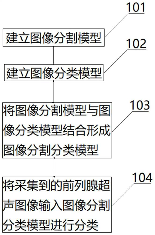
[0047] A method for segmenting and classifying prostate ultrasound images provided by an embodiment of the present invention includes the following steps:

[0048] 101. Establish an image segmentation model;

[0049] 102. Establish an image classification model;

[0050] 103. Combining the image segmentation model with the image classification model to form an image segmentation classification model;

[0051] 104. Input the collected ultrasound images of the prostate into the image segmentation classification model for classification.

[0052] Wherein, the steps of establishing an image segmentation model specifically include:

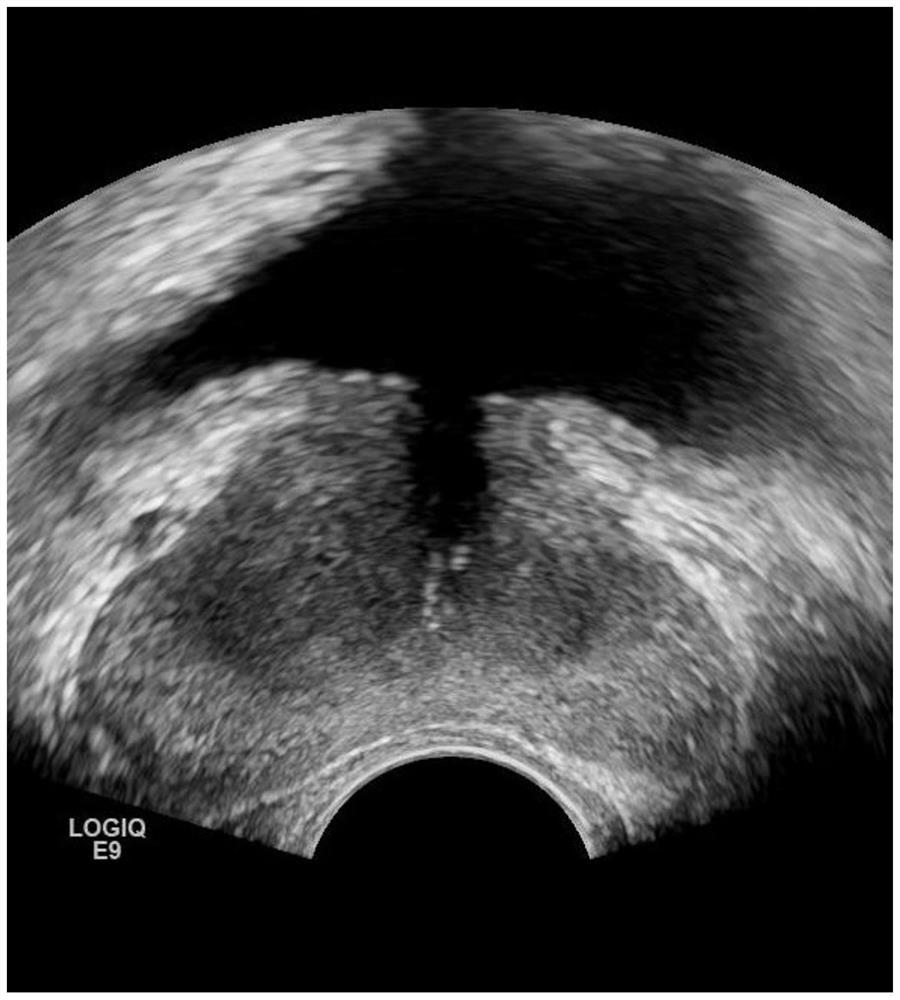
[0053] Use labelme software to segment the prostate region in the collected prostate ultrasound images to generate a manually segmented Mask map; establi...


PUM


Abstract

The invention discloses a prostate ultrasonic image segmentation and classification method. The method comprises the following steps: establishing an image segmentation model; establishing an image classification model; combining the image segmentation model with the image classification model to form an image segmentation classification model; and inputting the acquired prostate ultrasonic imagesinto an image segmentation and classification model for classification. Compared with the prior art, the method can improve the classification precision of the image, and improves the later diagnosisefficiency and accuracy.

Description

technical field [0001] The invention relates to the technical field of image processing, in particular to a method for segmenting and classifying prostate ultrasound images. Background technique [0002] Medical image classification is an important research direction in the field of computer vision. Classification technology provides application value for detection and disease treatment in the medical field. With the rapid development of artificial intelligence in the medical field, image classification has been extensively studied in the field of pattern recognition. and use. [0003] Due to serious problems such as speckle noise and low signal-to-noise ratio in medical image display, the classification of current medical images is not accurate enough and the degree of complexity is high, which brings a lot of trouble and pressure to doctors' later judgment and work. [0004] Therefore, how to provide an ultrasound image segmentation and classification method that can impr...

Claims


Application Information

Patent Timeline
Patent Type & Authority Applications(China)
IPC IPC(8): G06T7/11G06K9/62G06K9/32G06N3/04G06N3/08
CPCG06T7/11G06N3/08G06T2207/10132G06T2207/20081G06T2207/20084G06T2207/30081G06V10/25G06V2201/03G06N3/045G06F18/241G06F18/214
Inventor 卢旭沈顺博黄君卓育敏杨川
Owner GUANGDONG POLYTECHNIC NORMAL UNIV
Why Eureka
Try Eureka