

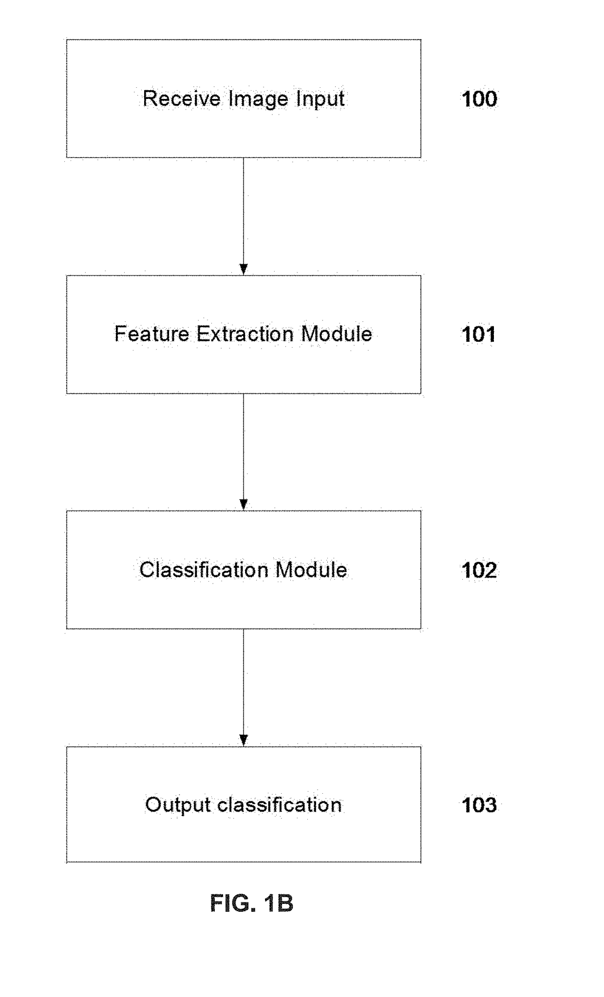
Classifying nuclei in histology images
a nucleus and image technology, applied in image analysis, instruments, computing, etc., can solve the problems of difficult, if not impossible, manual assessment of images by pathologists, and achieve superior classification results, improve nucleus classification accuracy, and improve the effect of nucleus classification
- Summary
- Abstract
- Description
- Claims
- Application Information
AI Technical Summary
Benefits of technology
Problems solved by technology
Method used
Image

Examples
example 1
[0142]A comprehensive evaluation was performed using a nucleus database with the nuclei detected from tumor-burdened tissue from PD-L1-stained lung samples. Slides were scanned on VENTANA iScan HT scanners, resulting in RGB whole slide images with 0.465 μm pixel size.
[0143]For these PD-L1-stained tissue images (see FIGS. 7A and 7B), five different types of nuclei needed to be classified. The PD-L1 database included 256 images of 600×700 pixels, which were obtained from PD-LI-stained lung samples. The slides were again scanned on VENTANA iScan HT scanners, resulting in RGB whole slide images with 0.465 μm pixel size. From these images, a number of nuclei of five types were selected by a pathologist, including nuclei from PD-LI-positive tumor cells (2,414), from PD-LI-negative tumor cells (1,620), from PD-LI-positive lymphocytes (2,851), from non-target staining cells (1,753), and from the remaining PD-LI-negative cells (1,834) (FIGS. 7A and 7B). These are PD-L1-positive tumor nuclei,...
example 2
[0148]An example for the scoring of a PD-L1-stained slide is shown in FIGS. 11A-D. The digital image of a tissue specimen immunohistochemically stained with PD-L1 (labeled with DAB in brown) and the counterstain hematoxylin is shown in FIGS. 11A and 11B. An automated analysis implementing the disclosed method has detected cells individually (not seen in this resolution) and labeled them as one of a PD-L1-positive tumor cell, a PD-L1-negative tumor cell, a PD-L1-positive lymphocyte, or any other cell. From these automatically generated read-outs, the tissue on this slide was scored individually for its PD-L1 status with respect to immune and immune cells. To score the tumor cells, the fraction of PD-L1-positive tumor cells is divided by the total number of tumor cells (i.e., PD-L1 positive and PD-L1 negative tumor cells). The tissue on this slide was scored as about 90% of the tumor cells being positive for PD-L1. To score the immune cells, the area in the image that contains these c...
PUM
 Abstract
Description
Claims
Application Information