
Pericardium puncture hydrops pumping device for cardiology department

A technique of pericardiocentesis and cardiology, applied in puncture needles, applications, medical science, etc., can solve the problems of not being too deep in the puncture site, difficult to grasp, and endangering the health of the operator, and achieve the effect of improving the bonding efficiency

Active Publication Date: 2020-10-13
NANYANG CITY CENT HOSPITAL
View PDF16 Cites 10 Cited by
  • Summary
  • Abstract
  • Description
  • Claims
  • Application Information

AI Technical Summary

Problems solved by technology

[0002] The Department of Cardiology, that is, the Department of Cardiovascular Medicine, is a clinical department set up by the major internal medicine departments of hospitals at all levels for the diagnosis and treatment of cardiovascular and vascular diseases. The diseases treated include angina pectoris, hypertension, sudden death, arrhythmia, heart failure, premature beats, arrhythmia, Myocardial infarction, cardiomyopathy, myocarditis, acute myocardial infarction and other cardiovascular diseases; pericardial effusion is a common cardiology disease. At present, the treatment of pericardial effusion is mainly to directly puncture the pericardial cavity with the help of a puncture needle and remove the effusion. Pull out, and the puncture site should not be too deep, so as not to puncture the atrium, ventricle or puncture the coronary artery and cause a large amount of hemorrhage in the pericardial cavity. Because the heart is constantly beating during treatment, it is difficult to master the puncture needle into the pericardial cavity. The puncture depth is extremely important. The existing liquid pumping device cannot extract the precise amount of liquid due to manual pumping, which brings inconvenience to the doctor's operation and is likely to endanger the health and even the life of the patient.

Method used


Image

  • Pericardium puncture hydrops pumping device for cardiology department
  • Pericardium puncture hydrops pumping device for cardiology department
  • Pericardium puncture hydrops pumping device for cardiology department

Examples


Embodiment Construction

[0029] The present invention will be further described below in conjunction with the accompanying drawings.

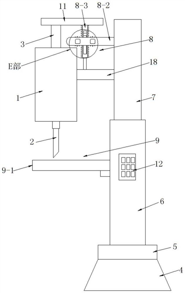
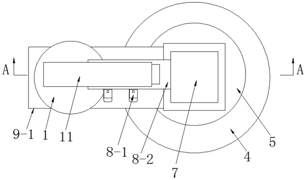
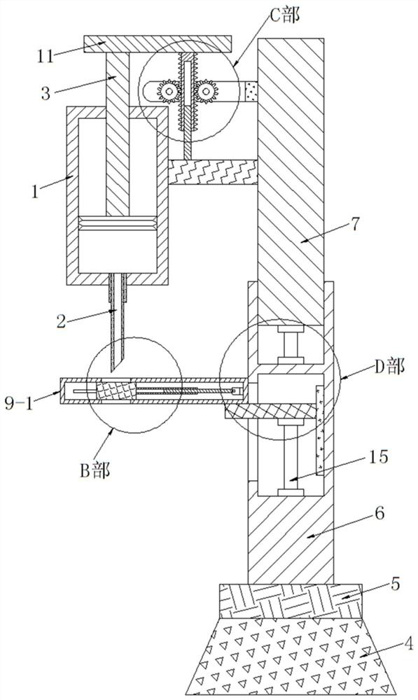
[0030] see as Figure 1-Figure 9 As shown, the technical solution adopted in this specific embodiment is: it includes a liquid collection tube 1, a puncture needle 2, and a push rod 3. The bottom of the liquid collection tube 1 is embedded and fixed with an adhesive puncture needle 2, and the collection A push rod 3 is inserted into the liquid cylinder 1, and the bottom pushing end of the push rod 3 is glued and fixed on the piston with an adhesive. The upper end of the push rod 3 is exposed on the upper side of the liquid collection tube 1; it also includes a suction cup 4, a base 5, a support 6, a support block 7, a pushing mechanism 8, and a chest protection mechanism 9. The suction cup 4 is fixed on the bottom of the base 5 with screws. The suction cup 4 is a vacuum suction cup. The device is adsorbed on the operating table through the suction cup 4. A support 6 i...


PUM


Abstract

The invention discloses a pericardium puncture hydrops pumping device for a cardiology department, and relates to the technical field of medical equipment. A suction cup is fixed to the bottom of a base; a support is fixed to the base; a chest protection mechanism is arranged on the left side of the support and arranged on the lower side of a puncture needle; a first groove is formed in the upperend of the support; a first air cylinder is fixed in the first groove; a supporting block is fixed to the pushing end of the upper end of the first air cylinder; a connecting block is fixed to the left side of the supporting block; the left side of the connecting block is fixedly connected with a liquid collecting barrel; an ejection block is fixed to the upper end of a push rod; and a pushing mechanism is arranged between the ejection block and the supporting block. A check block is additionally arranged at the chest position of a patient, so that the puncture needle is conveniently calibrated, and the subsequent puncture depth of the puncture needle is conveniently controlled; and moreover, the hydrops pumping amount is electrically controlled, so that the working intensity of a medicalstaff is relieved, and the hydrops pumping efficiency is improved.

Description

technical field [0001] The invention relates to the technical field of medical equipment, in particular to a pericardial puncture pumping device for cardiology. Background technique [0002] The Department of Cardiology, that is, the Department of Cardiovascular Medicine, is a clinical department set up by the major internal medicine departments of hospitals at all levels for the diagnosis and treatment of cardiovascular and vascular diseases. The diseases treated include angina pectoris, hypertension, sudden death, arrhythmia, heart failure, premature beats, arrhythmia, Myocardial infarction, cardiomyopathy, myocarditis, acute myocardial infarction and other cardiovascular diseases; pericardial effusion is a common cardiology disease. At present, the treatment method of pericardial effusion is mainly to directly puncture the pericardial cavity with the help of a puncture needle and remove the effusion. Pull out, and the puncture site should not be too deep, so as not to pun...

Claims


Application Information

Patent Timeline
IPC IPC(8): A61B17/34A61M1/00A61B90/00
CPCA61M1/0023A61B17/3403A61B90/08A61B17/3494A61B2017/3409A61M1/67
Inventor 王星
Owner NANYANG CITY CENT HOSPITAL
Learn More