
Three-dimensional cell and tissue image analysis for cellular and sub-cellular morphological modeling and classification

A subcellular and cellular technology, used in image analysis, graphic image conversion, image enhancement, etc.

Pending Publication Date: 2020-11-03
RGT UNIV OF MICHIGAN
View PDF0 Cites 2 Cited by
  • Summary
  • Abstract
  • Description
  • Claims
  • Application Information

AI Technical Summary

Problems solved by technology

However, the dimensionality and quality of imaging data and the great variability of cellular and subcellular shapes across populations pose challenges for 3D shape methods that should allow accurate morphological analysis, should be scalable , robust to noise and at the same time sufficiently specific across populations

Method used


Image

  • Three-dimensional cell and tissue image analysis for cellular and sub-cellular morphological modeling and classification
  • Three-dimensional cell and tissue image analysis for cellular and sub-cellular morphological modeling and classification
  • Three-dimensional cell and tissue image analysis for cellular and sub-cellular morphological modeling and classification

Examples


Embodiment Construction

[0028] Example embodiments will now be described more fully with reference to the accompanying drawings.

[0029] Tissue and cell morphology are regulated by complex underlying biological mechanisms related to cellular differentiation, development, proliferation, and disease. Changes in nuclear form reflect reorganization of chromatin structure associated with altered functional properties such as gene regulation and expression. At the same time, many studies in mechanobiology have shown that geometric constraints and mechanical forces act on cellular deformities and in turn affect nuclear and chromatin dynamics, gene and pathway activation. Thus, with the advent of the study of the reorganization of chromatin and DNA structures in spatial and temporal frames, known as the 4D nucleome, the morphological quantification of cells or organelles and components has become of interest. important meaning. Cellular structures of interest in the context of the 4D nucleome include not ...


PUM


Abstract

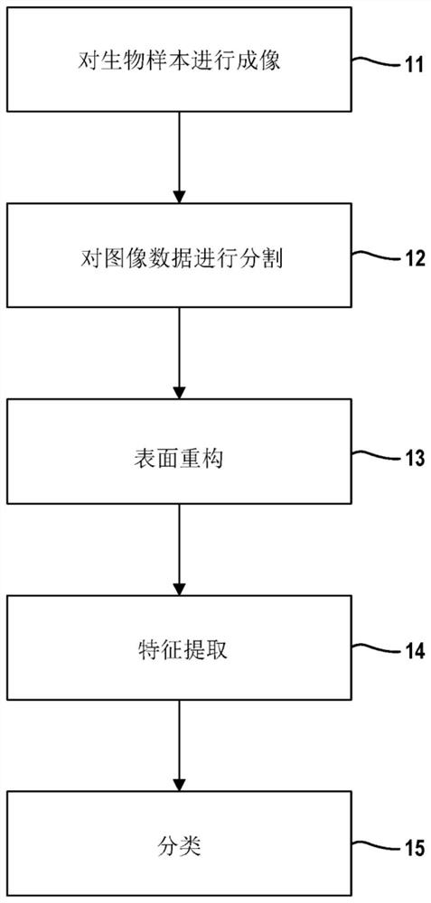
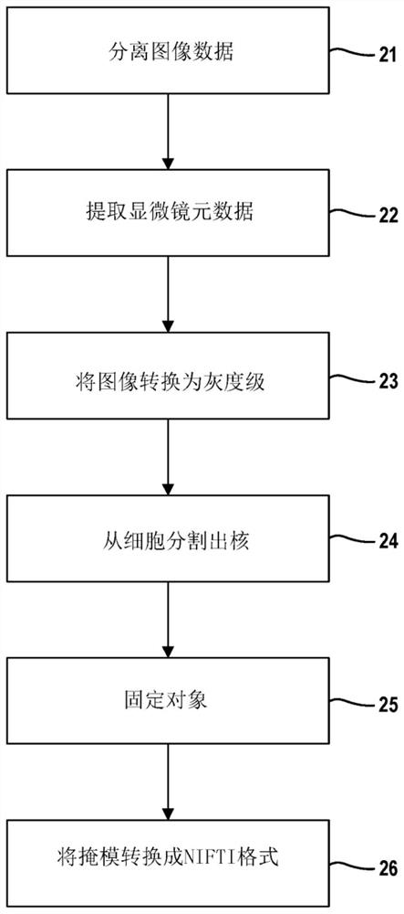
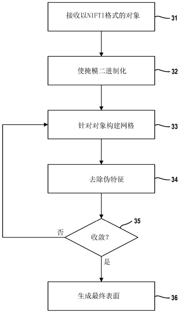
The ability to automate the processes of specimen collection, image acquisition, data pre-processing, computation of derived biomarkers, modeling, classification and analysis can significantly impactclinical decision-making and fundamental investigation of cell deformation. This disclosure combine 3D cell nuclear shape modeling by robust smooth surface reconstruction and extraction of shape morphometry measure into a highly parallel pipeline workflow protocol for end-to-end morphological analysis of thousands of nuclei and nucleoli in 3D. This approach allows efficient and informative evaluation of cell shapes in the imaging data and represents a reproducible technique that can be validated, modified, and repurposed by the biomedical community. This facilitates result reproducibility, collaborative method validation, and broad knowledge dissemination.

Description

[0001] government terms [0002] This invention was made with government support under NR015331, NS091856; DK089503 and EB020406 awarded by the National Institutes of Health, and IIS-636840 awarded by the National Science Foundation. The government has certain rights in this invention. [0003] Cross References to Related Applications [0004] This application claims priority to U.S. Patent Application No. 16 / 277,128, filed February 15, 2019, and also claims the benefit of U.S. Provisional Application No. 62 / 632,663, filed February 20, 2018. The entire disclosure of the above application is incorporated herein by reference. technical field [0005] The present disclosure relates to three-dimensional cell and tissue image analysis for modeling and classification of cellular and subcellular morphology. Background technique [0006] Tissue and cellular deformation is regulated by complex underlying biological mechanisms that affect spatial and temporal morphological changes....

Claims


Application Information

Patent Timeline
Patent Type & Authority Applications(China)
IPC IPC(8): G06T17/20G06T7/11G06T3/40G16B45/00
CPCG01N1/30G06T7/12G06T7/143G06T7/0012G06T2207/30024G06T2207/10056G06T2207/30242G06T2207/20084G06T2207/10064G06T2207/10101G06T2207/20072G06T2207/20036G06T2207/20081G06V20/695G06V20/698G06V20/64G06T7/62G06T7/50G06F18/24765G06F18/2411G06F18/2415G06F18/2451G06F18/24147
Inventor 伊瓦伊洛·迪诺夫布赖恩·D·阿西大卫·S·迪尔沃思阿里·阿林-福耶尔亚历山大·加里宁亚历克斯·S·阿德
Owner RGT UNIV OF MICHIGAN
Learn More